Fig. S2
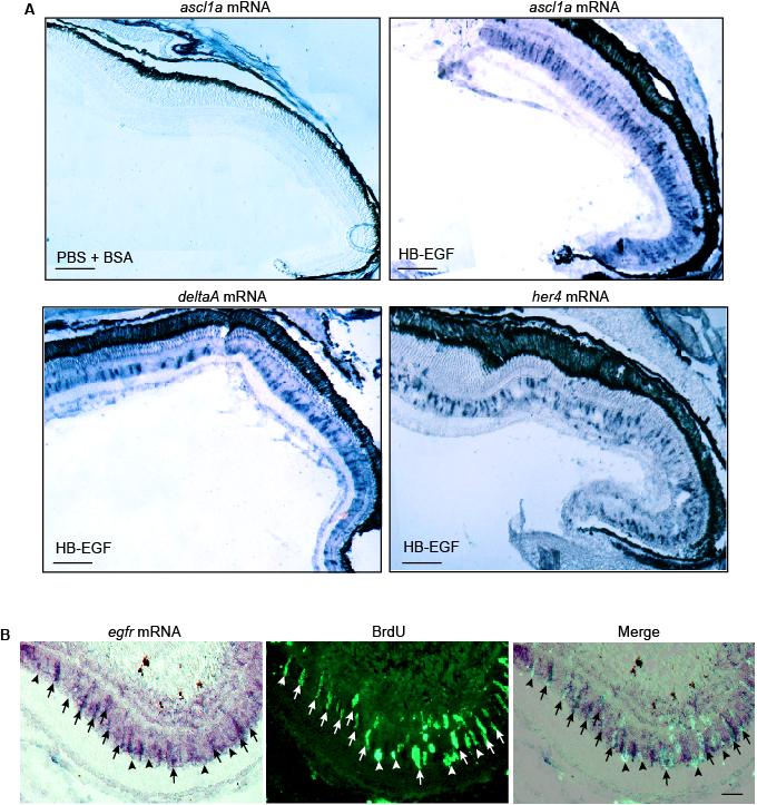
HB-EGF stimulates ascl1a, deltaA and her4 gene expression throughout the uninjured retina. Related to Figure 2. (A) Low magnification view of in situ hybridization assays showing that intravitreal injection of HB-EGF stimulates ascl1a, deltaA and her4 gene expression throughout the retina. In situ hybridization assays for control eyes that only received PBS + BSA showed no expression above background (data shown for ascl1a mRNA, which is representative of the other mRNAs). Scale bar, 150 μm. (B) In situ hybridization and BrdU immunofluorescence shows EGFR mRNA is localized to proliferating MG at the injury site. Arrows identify proliferating MG with strong EGFR expression while arrowheads point to proliferating MG with weak EGFR expression. Scale bar, 50 μm.
Reprinted from Developmental Cell, 22(2), Wan, J., Ramachandran, R., and Goldman, D., HB-EGF Is Necessary and Sufficient for Müller Glia Dedifferentiation and Retina Regeneration, 334-347, Copyright (2012) with permission from Elsevier. Full text @ Dev. Cell

