
|
Fig. 6
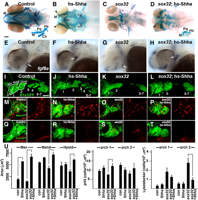
Shha misexpression partially rescues the jaw defects of sox32 mutants. (A–D) Ventral views of 6 dpf head skeletons. Compared to hsp70I:Gal4 controls subjected to the same heat-shock treatment (A), hs-Shha larvae developed ectopic Tr-like cartilages (arrowheads) (B, n = 10/10). Whereas facial skeleton never formed in non-hs-Shha sox32 siblings (C, n = 0/10), jaw and hyoid cartilage was partially restored and ectopic cartilage was still present in sox32; hs-Shha larvae (D, n = 4/4). Flat-mount preparations (insets in A and D) show that Meckel′s (M), palatoquadrate (Pq), and hyosymplectic (Hs) cartilages, but not the hyoid-arch-derived ceratohyal (Ch) cartilage, were dysmorphic yet present in sox32; hs-Shha larvae. (E–H) In situ hybridizations show fgf8a oral ectoderm expression (arrows) at 36 hpf. Compared to hsp70I:Gal4 controls (E), fgf8 expression was shifted dorsal-anteriorly in hs-Shha embryos (F, n = 8/9) and partially (n = 3/34) or severely reduced (n = 31/34) in sox32 mutants (G). Shha misexpression resulted in increased and dorsal-laterally shifted fgf8a expression in sox32; hs-Shha embryos (H, n = 11/12). (I–L) Confocal projections show the pharyngeal arches (numbered) of control fli1a:GFP (I, n = 3), hs-Shha; fli1a:GFP (J, n = 4), sox32; fli1a:GFP (K, n = 10), and sox32; hs-Shha; fli1a:GFP (L, n = 7) embryos at 30 hpf. In sox32 and sox32; hs-Shha embryos, the lack of endoderm resulted in arches 3–7 remaining as a single mass. White outlines show the maxillary (max), mandibular (mand), and hyoid domains for wild type. (M–T) Anti-pH3 staining marks proliferating cells (red, M–P), Lysotracker marks regions of cell death (red, Q–T), and fli1a:GFP labels CNCCs (green) of the mandibular (1) and hyoid (2) arches in control (M and Q), hs-Shha (N and R), sox32 (O and S), and sox32; hs-Shha (P and T) embryos at 36 hpf. n = 10 for each genotype. Scale bars = 50 μm. (U) Quantification of arch domain area and numbers of pH3- and Lysotracker-positive cells per arch area in control hsp70I:Gal4 (con), hs-Shha, sox32, and sox32; hs-Shha embryos. Brackets indicate comparisons showing statistical significance with a Tukey–Kramer HSD test (α = 0.05).
Reprinted from Developmental Biology, 361(2), Balczerski, B., Matsutani, M., Castillo, P., Osborne, N., Stainier, D.Y., and Crump, J.G., Analysis of Sphingosine-1-phosphate signaling mutants reveals endodermal requirements for the growth but not dorsoventral patterning of jaw skeletal precursors, 230-241, Copyright (2012) with permission from Elsevier. Full text @ Dev. Biol.