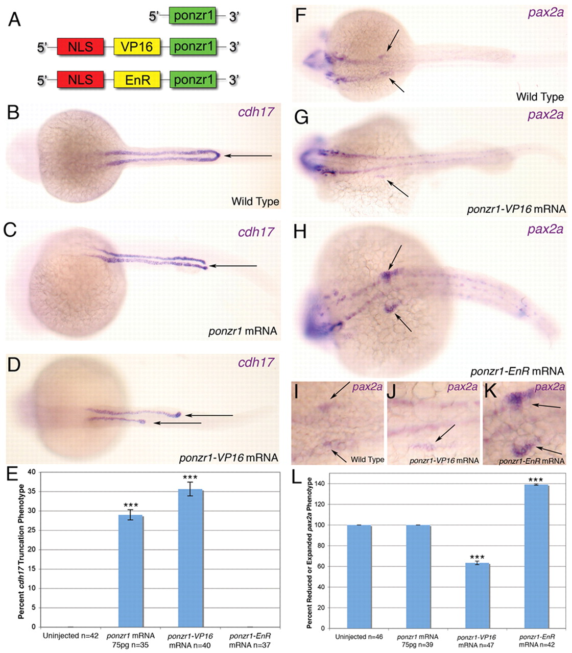
Fig. 8
ponzr1 can function as a transcription factor or co-factor. (A) Diagrams showing the transcriptional activator and repressor mRNAs injected into zebrafish. The NLS was included to facilitate nuclear access of the fusion proteins. (B) Dorsal view of wild-type cdh17 expression in the pronephric ducts at 24 hpf. (C) ponzr1 mRNA-injected embryos reveal cdh17 expression loss at the cloaca (arrow). (D) ponzr1-VP16 mRNA-injected embryos show a severely truncated cdh17 expression in the pronephric ducts (arrows). (E) cdh17 phenotype in ponzr1- and ponzr1-VP16-injected embryos are significantly different from control, whereas ponzr1-EnR-injected embryos are not. (F) Wild-type pax2a expression in the anterior pronephric ducts at 24 hpf (magnified I). (G-K) ponzr1-VP16 mRNA-injected embryos (G) show reduced pax2a expression (magnified J) whereas ponzr1-EnR mRNA-injected embryos (H) demonstrate expanded pax2a expression in the anterior pronephric ducts (arrows) (magnified K) at 24 hpf. (L) Quantification of pax2a phenotypes standardized to wild-type pax2a at 100%. Percentage of embryos with reduced pax2a is seen as below 100% (as seen in ponzr1VP16-injected embryos) and expanded pax2a expression is seen above 100% (as seen in ponzr1EnR-injected embryos). Both ponzr1-VP16 and ponzr1-EnR are significantly different from wild type. ***P<0.001

