
|
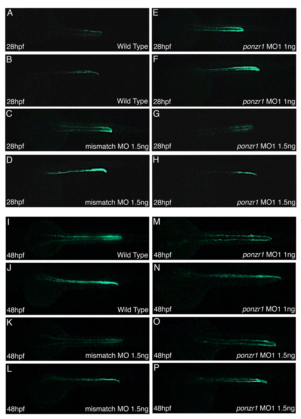
Fig. S3
Pronephric tubule and duct marker Tg(atp1a1a.4:GFP) shows no changes in pronephric patterning in ponzr1 morphants. (A-P) Wild-type Tg(atp1a1a.4:GFP) embryos at 28 hpf (A, dorsal; B, lateral) and 48 hpf (I, dorsal; J, lateral) show fluorescence in the pronephric tubules and ducts. Mismatch MO-injected embryos show normal fluorescence patterns at 28 hpf (C, dorsal; D, lateral) and 48 hpf (K, dorsal; L, lateral). Embryos were injected at 1 ng (E,F) and 1.5 ng (G,H) of ponzr1 MO1 show no differences in the amount and localization of the fluorescence at 28 hpf (E,G, dorsal; F,H, lateral) and 48 hpf (M,O, dorsal; N,P, lateral). This experiment was conducted twice viewing at least 35 embryos for each group.