
|
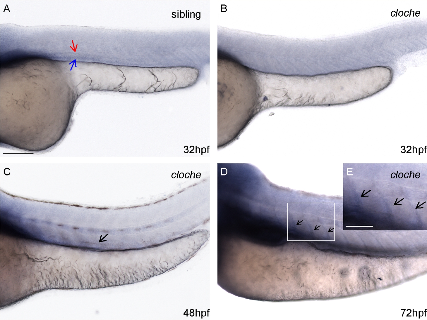
Fig. S2
Prox1b expression in cloche mutant. (A?D) shows prox1b expression in cloche mutants and siblings. The red and blue arrows point to prox1b expression in the DA and PCV of a sibling embryo separately (A), while this prox1b expression is absent in cloche mutant (B). The black arrows point to signals in the ventral region of the trunk (C) and along the somite boundaries of homozygous cloche mutants (D). (E) The inset shows an enlarged view of the boxed area in (D). The staining indicated by black arrows in C, D and E is either non-endothelial or non-specific because cloche mutant embryos lack endothelial cells. Given the diffuse nature of the staining, the black arrow indicated signal is probably a non-specific artifact of over-staining. Scale bars represent 100 μm in (A), and 50 μm in (E).