
|
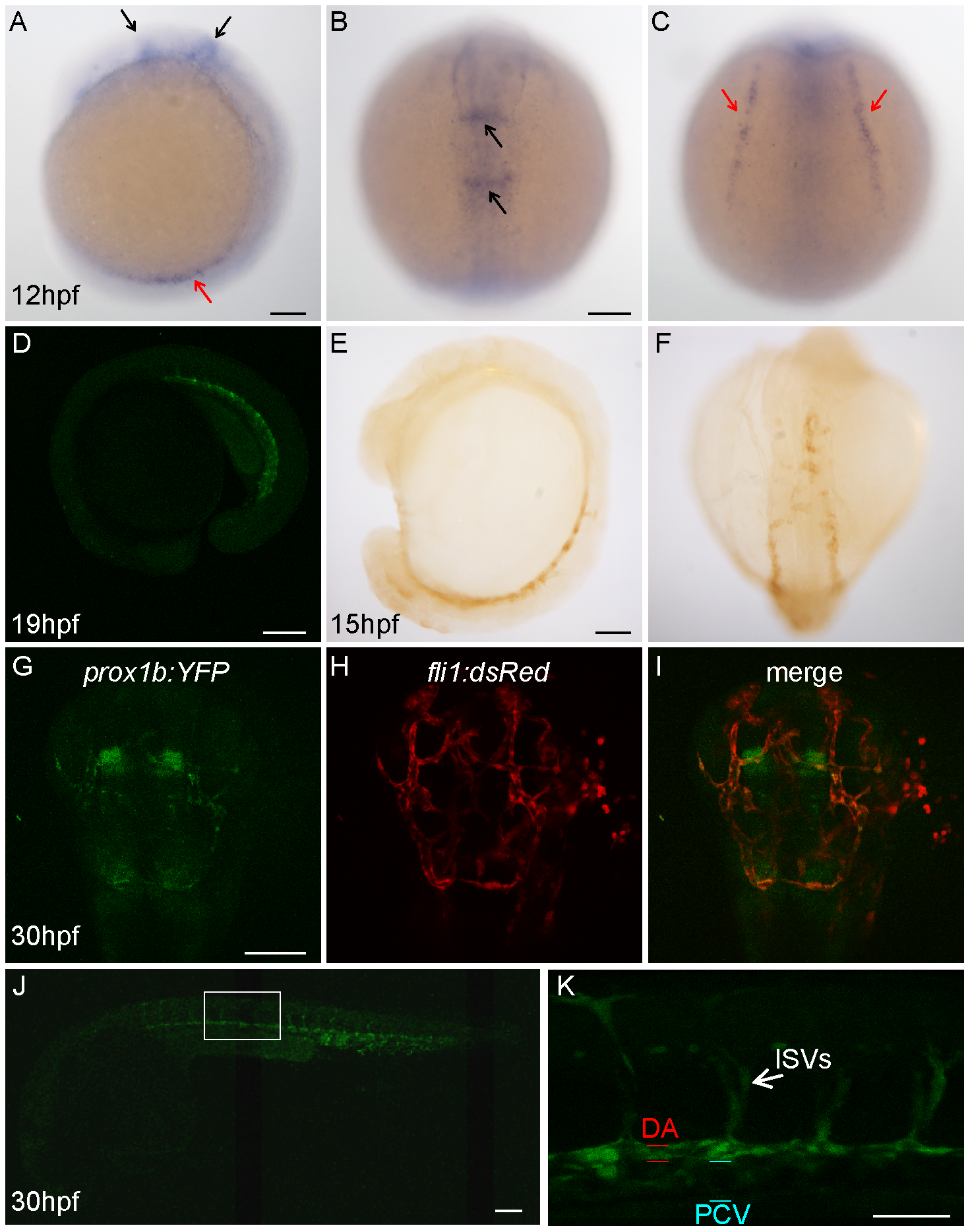
Fig. 1
Prox1b is expressed in the endothelial cells and the central nervous system of the head.
(A–C) shows prox1b transcript expression by whole mount in situ hybridization in wild-type embryos, at 12 hpf. Black arrows point to prox1b expression in the head; red arrows indicate prox1b expression in lateral plate mesoderm. Confocal image (D) shows YFP expression in a prox1b BAC:YFP embryo at 19 hpf stage. (E) and (F) show YFP expression, enhanced by DAB immunostaining, is detected in prox1b BAC:YFP embryos in migrating angioblasts at 15 hpf. (G–I) shows prox1b:YFP expression in the head region of a prox1b BAC:YFP, fli1:DsRed embryo. Note overlapping (endothelial cells) and non-overlapping expression domains. (J) shows prox1b:YFP expression in the trunk vasculature. (K) shows enlarged view of the boxed area in (J). Scale bars represent 50 μm in (K), and 100 μm in other figures.