
Image
|
Figure Caption
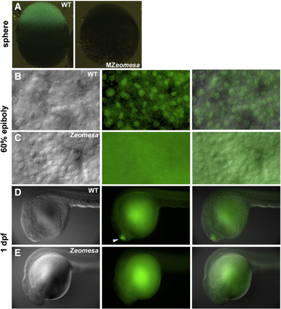
Fig. S2 Eomesa in MZeomesa and Zeomesa embryos. Overlay of fluorescence and bright field images of (A) wild type and (B) MZeomesa embryos stained for Eomesa at sphere stage. No nuclear or cytoplasmic fluorescence is detected in MZeomesa embryo. (B-E) Bright field, fluorescene and overlay of embryos stained for Eomesa. Stages indicated on left, genotype in upper right. (B,C) Nuclear staining visible in wild type deep cells but not Zeomesa deep cells at 60% epiboly. Background fluorescence is visible in mutant embryo. (D) Eomesa detected in the brain of wild type (arrowhead) but not Zeomesa (E) embryos at 1 dpf.