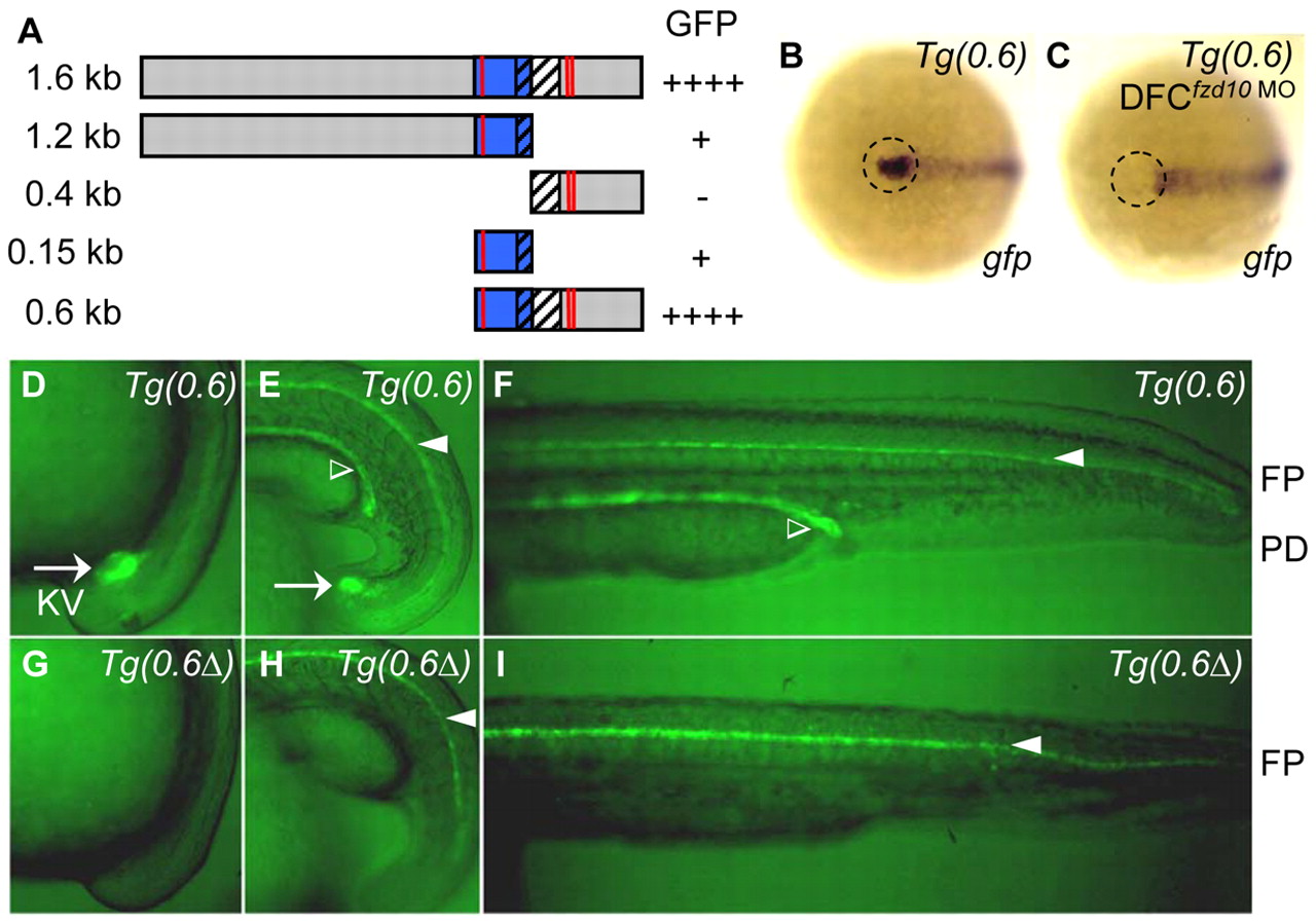
Fig. 5 Wnt signaling directly regulates foxj1a transcription. (A) Schematic of foxj1a enhancers. Sequence of foxj1a that is located approximately –6.2 kb to –4.6 kb upstream of the ATG start codon was used to generate a series of report constructs. Blue bar, conserved sequence between zebrafish and tetradon. Hatched bar, predicted first non-coding exon. Red line, putative Lef1/Tcf binding site. These fragments were inserted in front of the gfp sequence in a tol2 vector, and the resulting plasmids were co-injected with tol2 transposase RNA into embryos. Fluorescent GFP signals in KV were scored. (B,C) A 0.6 kb enhancer sequence contains Wnt-responsive cis-acting elements. Transgenic Tg(0.6foxj1a:gfp) embryos showed reporter gfp expression in DFCs (B; 12/12). DFC-targeted injection of fzd10 MO abolished gfp expression in DFCs (C; 14/15). Shown is in situ hybridization using gfp as a probe in a ventral view of 95% epiboly staged embryos. Dashed circle indicates DFC region. (D-I) Putative Lef1/Tcf binding sites are required for foxj1a expression in KV. GFP reporter expression in stable transgenic Tg(0.6foxj1a:gfp) embryos recapitulated foxj1a expression pattern in KV (D,E), PDs (E,F) and FP (E,F). After all three putative Lef1/Tcf binding sites in the 0.6 kb enhancer were deleted using a site-directed mutagenesis kit, stable transgenic Tg(0.6Δfoxj1a:gfp) embryos lacked GFP expression in KV (G,H) and PDs (H,I) but maintained GFP expression in FP (H,I). Arrow indicates KV, open arrowhead PDs and filled arrowhead FP. Shown are embryos at 10-12 somites (D,G), 16 somites (E,H) and 30 hpf (F,I).
Image
Figure Caption
Figure Data
Acknowledgments
This image is the copyrighted work of the attributed author or publisher, and
ZFIN has permission only to display this image to its users.
Additional permissions should be obtained from the applicable author or publisher of the image.
Full text @ Development

