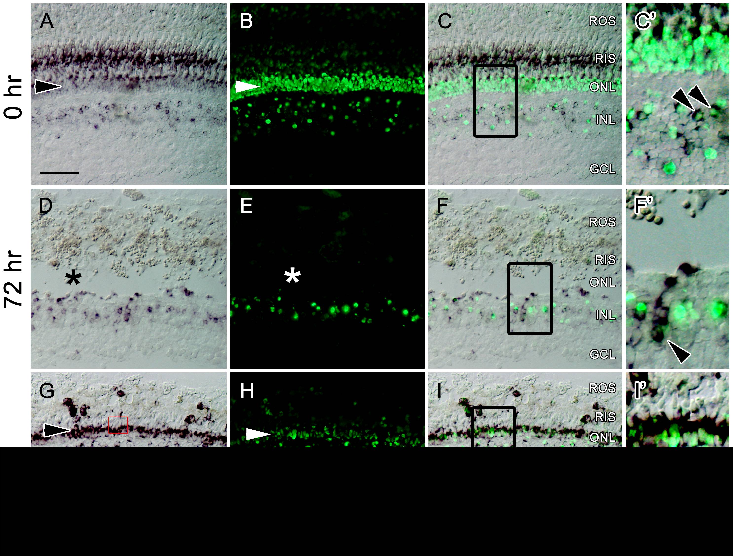
Fig. 11 RNA in situ hybridization on retinal sections from adult Tg(nrd:egfp)/albino zebrafish comparing endogenous neurod expression (purple) to transgene expression (green) during light-induced retinal regeneration.
(A) Before light treatment (0 hr), endogenous neurod is expressed in a subset of amacrine and bipolar cells, weakly expressed in rod photoreceptors in the ONL (arrowhead), and expressed in rod inner segments (RIS). (B) The nrd:egfp transgene is expressed in a subset of amacrine and bipolar cells, in rod photoreceptors in the ONL (arrowhead), and weakly in RIS. (C) Overlay of panels (A) and (B). (C2) Higher magnification inset of (C) showing co-labeling of endogenous and transgenic neurod expression in a subset of cells in the INL (arrowheads). (D) 72 hours after light onset (72 hr), all rod photoreceptors have been ablated (indicated by the asterisk), and endogenous neurod is persistently expressed in a subset of amacrine and bipolar cells. (E) The nrd:egfp transgene is persistently expressed in a subset of amacrine and bipolar cells. (F) Overlay of panels (D) and (E). (F′) Higher magnification inset of (F) showing co-labeling of endogenous and transgenic neurod expression in a column of progenitor cells (indicated by the arrowhead), and a subset of cells of the INL. (G) 7 days after light onset (7 d), endogenous neurod is strongly expressed in newly formed rods in the ONL (black arrowhead), and persistently expressed in a subset of amacrine and bipolar cells. The inset shows expression of neurod in newly-formed rod inner segments (white arrowhead). (H) The transgene is more weakly expressed in newly formed rods, and persistently expressed in a subset of amacrine and bipolar cells. (I) Overlay of panels (G) and (H). (I′) Higher magnification inset of (I) showing co-labeling of endogenous and transgenic neurod expression in a subset of INL cells (arrowhead), and in newly formed rod progenitors. Scale bar: 50 microns (A?C, D?F, G?I).

