
Image
|
Figure Caption
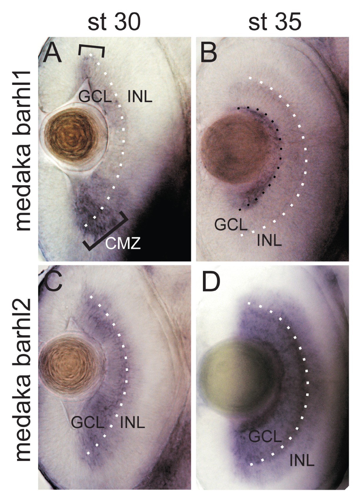
Fig. 8
Expression patterns of the medaka barhl1 and barhl2 in the retina. Dorsal views through the retina of medaka embryos hybridized with either medaka barhl1 (A, B) or medaka barhl2 (C, D) RNA antisense probe. Anterior is to the top. (A) Expression of medaka barhl1 restricted to the ciliary marginal zone (CMZ) is highlighted with black brackets in A and with a black dotted line in B. The white dotted line indicates the ganglion cell layer (GCL)/inner nuclear layer (INL) boundary.
Acknowledgments
This image is the copyrighted work of the attributed author or publisher, and
ZFIN has permission only to display this image to its users.
Additional permissions should be obtained from the applicable author or publisher of the image.
Full text @ BMC Evol. Biol.