
|
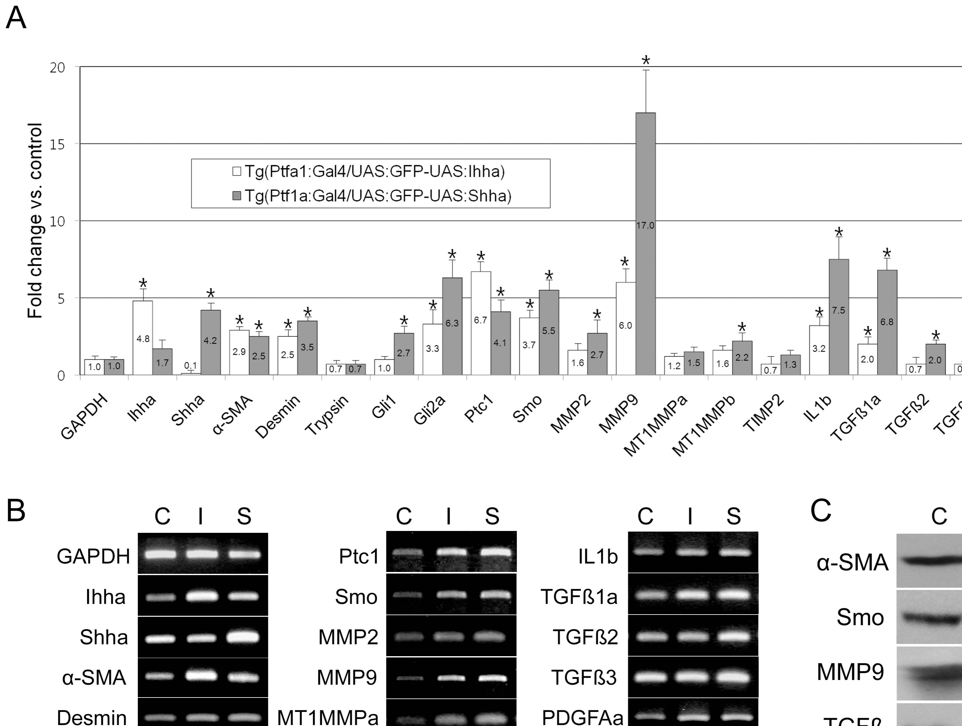
Fig. 5 RT-PCR and Western blot.
Pancreas from 3?4 month-old zebrafish was dissected under a fluorescence microscope. C, Tg(Ptf1a-Gal4/UAS:GFP); I, Tg(Ptf1a-Gal4/UAS:GFP-UAS:Ihha); S, Tg(Ptf1a-Gal4/UAS:GFP-UAS:Ihha). (A) Real-time RT-PCR showing differential expression of the components of the Hh pathway and fibrosis by Hh over-expression. Note the prominent up-regulation of MMP9 and TGFβ1a. (B) Electrophoretic images of RT-PCR products recapitulate real-time PCR data. (C) A western blot hybridization using available antibodies which are reactive to zebrafish antigens also recapitulates RT-PCR findings. α-SMA, 42 kD; Smo, 85 kD; MMP9, 75 kD; TGF�, 45 kD; β-actin, 45 kD. * P<0.05 versus control.