
|
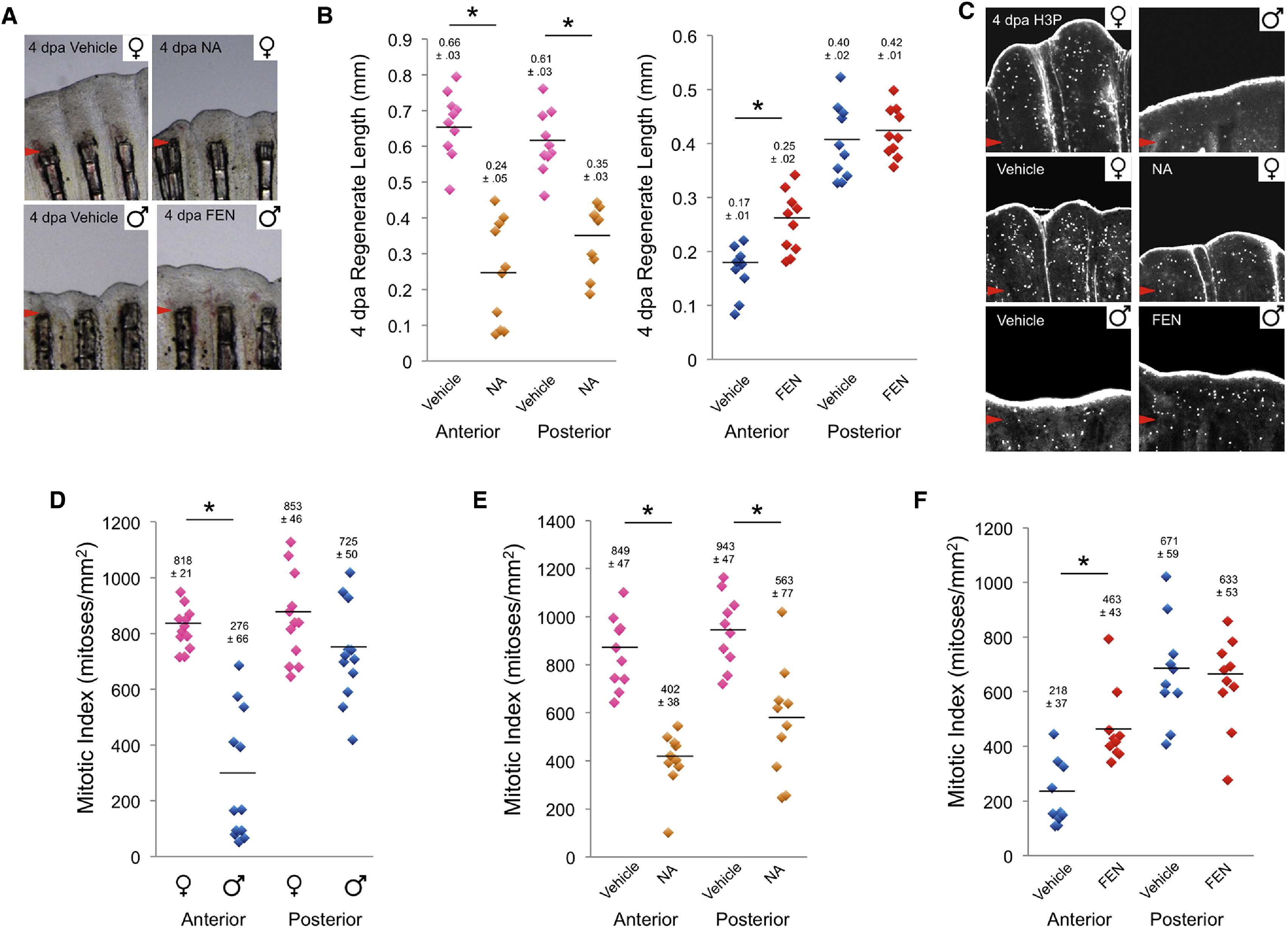
Fig. 2
Androgen Signaling Inhibits Blastemal Proliferation
(A) Top: 4 days of androgen treatment (1 μg/ml NA) after amputation inhibited pectoral fin regeneration in females. Bottom: androgen receptor antagonist treatment (1 μg/ml FEN) for 4 days after amputation improved male fin regeneration. Anterior regions are shown. Arrowheads indicate amputation plane.
(B) Quantification of effects of NA on female fin regeneration (left) (n = 10; *p < 0.001 by Student′s t test) and FEN on male fin regeneration (right) (n = 10; *p < 0.005 by Student′s t test).
(C) Top: blastemal proliferation in anterior portions of male and female 4 dpa pectoral fin regenerates, assessed by phosphorylated histone H3 (H3P) staining. Images are confocal projections of the medial 10 μm of mesenchyme; the anti-H3P antibody also nonspecifically stains the fin epidermis. Middle: blastemal proliferation in females treated with vehicle or NA. Bottom: blastemal proliferation in males treated with vehicle or FEN. Anterior regions are shown.
(D) Quantification of blastemal proliferation in females and males (n = 12). *p < 0.001 by Student′s t test.
(E) Quantification of effects of NA on female blastemal proliferation (n = 10). *p < 0.001 by Student′s t test.
(F) Quantification of effects of FEN on male blastemal proliferation (n = 10). *p < 0.001 by Student′s t test.