
|
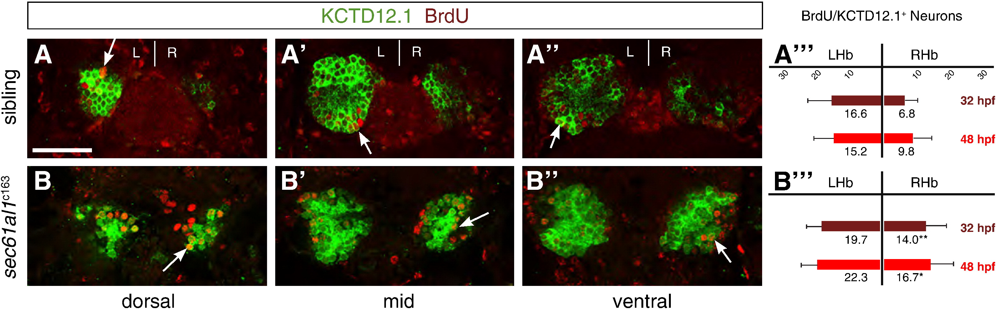
Fig. 6 Increased cell division of LsDh precursors in sec61al1c163 mutants. Embryos were briefly pulsed with BrdU at 32 hpf and chased until 4 dpf, when they were labeled for Kctd12.1 (green) and BrdU (red). As neurons are post-mitotic, brightly labeled BrdU+ cells are those that underwent a final mitosis during the BrdU pulse (white arrows); see text for further explanation of the labeling strategy. (A?A′′′) Compared to WT controls, (B?B′′′) significantly more neurons are born in the RHb of sec61al1c163 mutants at 32 hpf. This trend continues through 48 hpf (A′′′,B′′′), as again there are significantly more neurons in the RHb of sec61al1c163 mutants (images not shown) (Andres et al., 1999). Images are single z-plane images from confocal z-stacks. Dorsal (A,B), mid (A′,B′), and ventral (A′′,B′′) sections are shown. Scale bar represents 30 μm. Significance determined by Student′s T-test (*p < 0.05; **p < 0.01); n = 41 siblings, n = 43 mutants.
Reprinted from Developmental Biology, 360(1), Doll, C.A., Burkart, J.T., Hope, K.D., Halpern, M.E., and Gamse, J.T., Subnuclear development of the zebrafish habenular nuclei requires ER translocon function, 44-57, Copyright (2011) with permission from Elsevier. Full text @ Dev. Biol.