
|
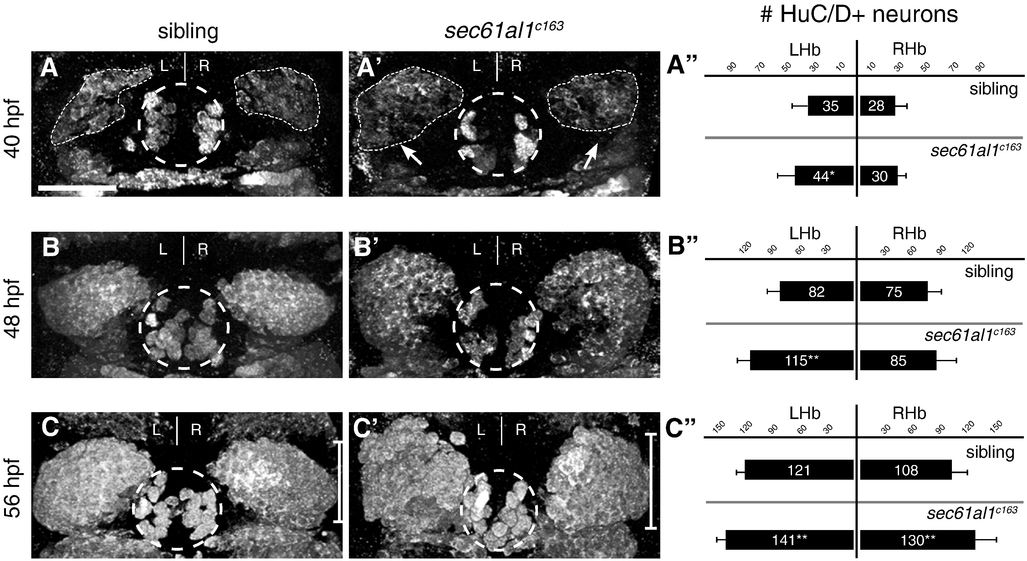
Fig. 5 Excess habenular neurons form in sec61al1c163 mutant habenulae. (A–C′) Post-mitotic neurons in WT or sec61al1c163 mutant embryos as detected by HuC/D antibody labeling. (A) A representative sibling embryo has fewer HuC/D+ habenular neurons at 40 hpf as compared to (A′) a sec61al1c163 mutant. (A") Quantification of data at 40 hpf. The number of left habenula neurons was significantly increased in homozygous mutants compared to WT siblings. (B–B") At 48 hpf, significantly more neurons were detected in the left habenula of mutants than in WT controls. (C–C′′) By 56 hpf, both the left and right habenulae of mutants contain more neurons than controls. In all panels, the pineal organ is outlined by a dashed circle. Scale bar equals 30 μm. Significance determined by Student′s T-Test (*p < 0.05, **p < 0.01); 40 hpf (n = 12), 48 hpf (n = 9), 56 hpf (n = 12). Images are extended focus projections of confocal Z-stacks through the dorsal diencephalon.
Reprinted from Developmental Biology, 360(1), Doll, C.A., Burkart, J.T., Hope, K.D., Halpern, M.E., and Gamse, J.T., Subnuclear development of the zebrafish habenular nuclei requires ER translocon function, 44-57, Copyright (2011) with permission from Elsevier. Full text @ Dev. Biol.