
|
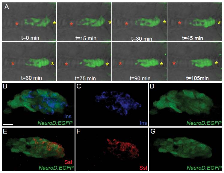
Fig. S3 NeuroD: enhanced green fluorescent protein (EGFP) cells are endocrine precursors. (A) TgBAC(NeuroD:EGFP)nl1 embryos, mounted dorsal side up, imaged by confocal timelapse microscopy starting at 19 h post fertilization (hpf). Images were captured every 15 min. Asterisks indicate fixed points of the embryo determined from the bright field image. Anterior is to the left. (B-G) Confocal image projection of 24 hpf TgBAC(NeuroD:EGFP)nl1 embryo immunostained for green fluorescent protein (GFP) and insulin (Ins) (B-D) and GFP and somatostatin (Sst) (E-G), showing overlap of NeuroD:EGFP expression with islet hormones in a subset of cells. All are ventral view. Scale bar = 15 μM.