
|
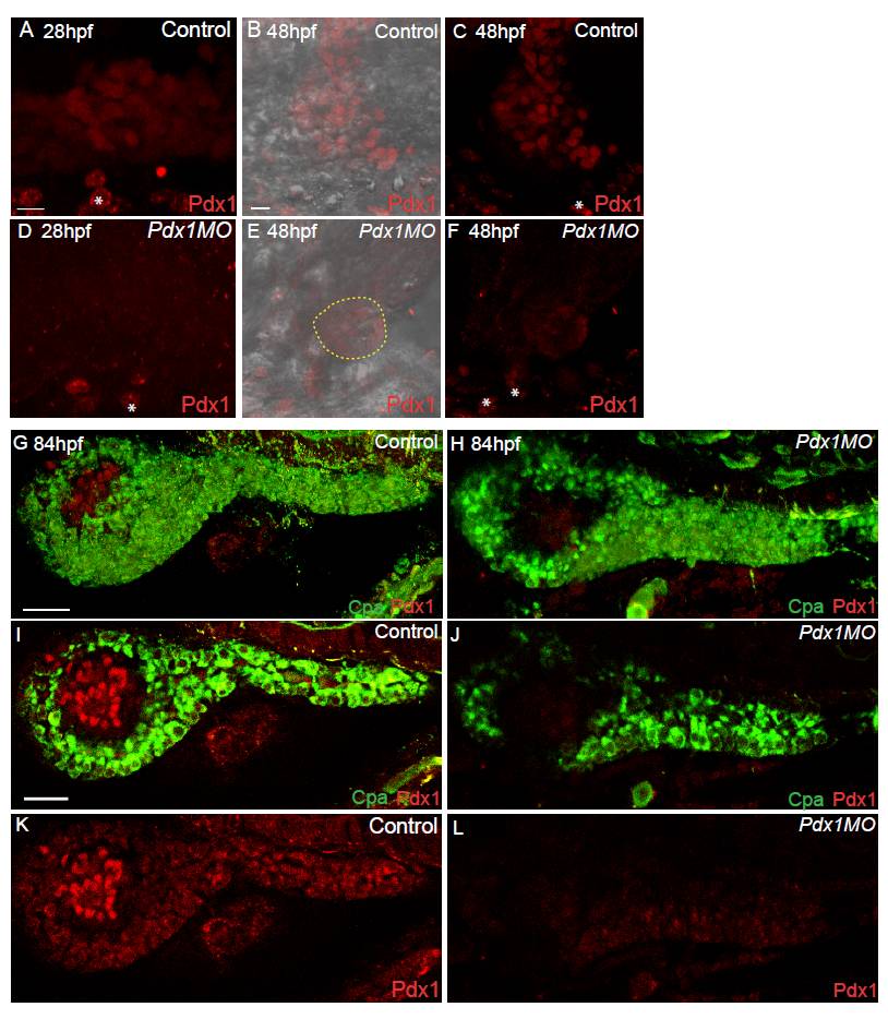
Fig. S2 Pdx1 protein expression in pdx1 morphants. Confocal projections of control and pdx1 morpholino-injected embryos at 28 h post fertilization (hpf) (A-C) and 48 hpf (D-F), immunostained for Pdx1. (A-C) Control embryos express Pdx1 in the developing gut and pancreas. (D-F) pdx1 morphant embryos have no detectable Pdx1 expression. (B,E) Overlay of bright field with confocal projection of Pdx1 antibody staining, (C,F) single channel showing Pdx1 antibody. Dashed circle (E) indicates islet as determined from bright field images. Non-specific labeling by this antibody of somites lateral to the gut (asterisks) has been previously described [49]. Ventral view. Scale bar = 10 μM. Confocal projections (G,H) and single plane views (I-L) of control and pdx1 morphants at 84 hpf, immunostained for Pdx1 and carboxypeptidase A (Cpa) to indicate the exocrine pancreas. Control embryos show robust Pdx1 expression in the islet and weaker expression in the exocrine pancreas (G,I,K). pdx1 morphant embryos have low-level Pdx1 expression in the islet and exocrine pancreas (H,J,L). Lateral view, anterior to left. Scale bar = 30 μM.