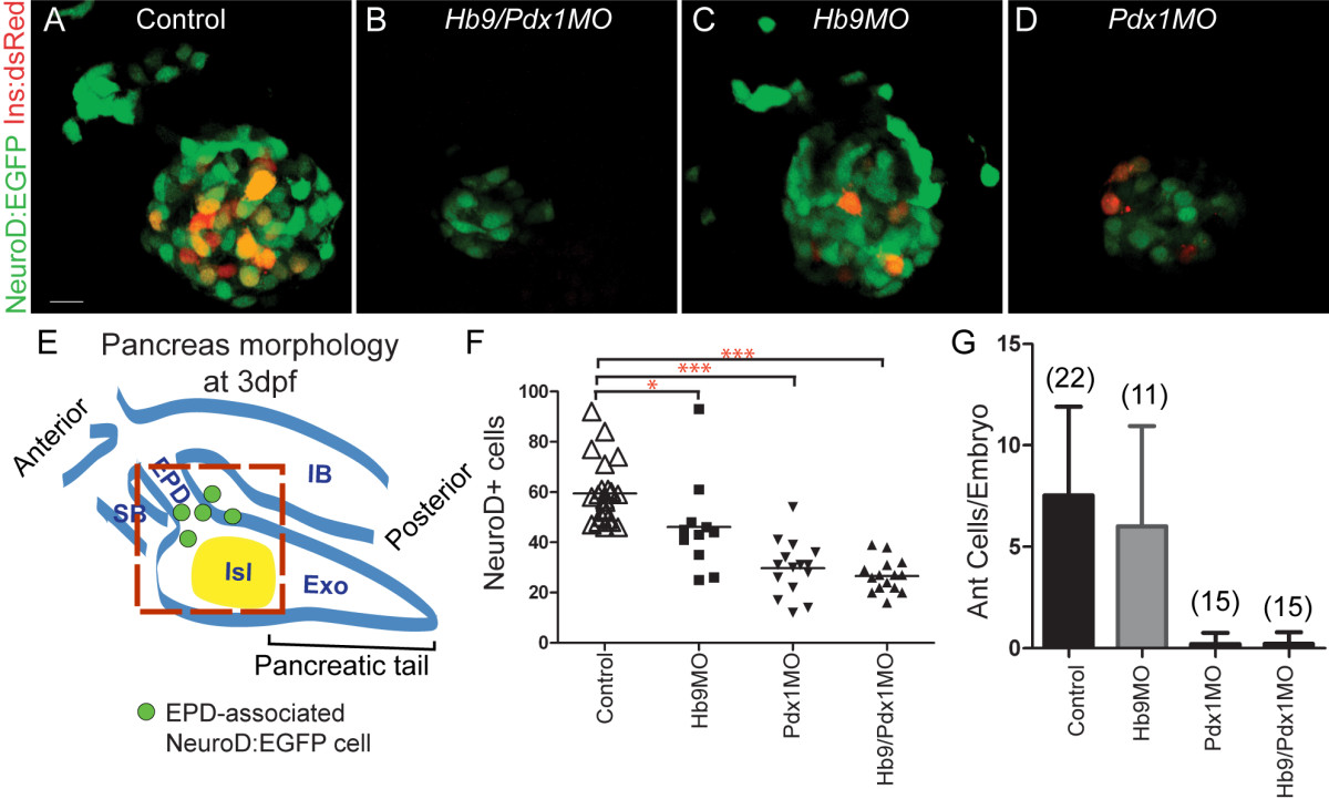
Fig. 3 Absence of ventral bud endocrine precursors in hb9/pdx1 and pdx1 morphant embryos. (A-D) Projections of confocal stacks showing native fluorescence of 3 days post fertilization (dpf) TgBAC(NeuroD:EGFP)nl1; Tg(ins:dsRed)m1018 embryos. In uninjected control embryos (A) and in hb9 morphants (C), enhanced green fluorescent protein (EGFP)+ cells are found in the islet and in smaller numbers also anterior to the islet. In hb9/pdx1 double morphants (B) and pdx1 morphants (D), the number of islet-associated EGFP+ cells is strongly reduced and anterior cells are missing. Few Ins:DsRed cells are present in pdx1 single morphants at 3 dpf (D), reflecting slow maturation of the DsRed fluorophore. (E) Schematic of pancreas morphology at 3 dpf. The principal islet (Isl) at this stage is primarily dorsal bud derived. The ventral bud contributes new NeuroD:EGFP cells (green circles), and generates exocrine pancreas (Exo) and extrapancreatic duct (EPD). IB, intestinal bulb; SB, swim bladder. Red box delineates region included for quantitation of islet and newly emerging anterior endocrine cells. (F) Quantitation of pancreatic EGFP+ cells (*P < 0.05, ***P < 0.001 with P values determined using one-way analysis of variance (ANOVA) with Bonferroni′s post test). (G) Quantitation of anteriorly positioned EGFP+ cells in uninjected and morpholino injected embryos (error bars indicate standard deviation from the mean). Scale bar = 10 μM.
Image
Figure Caption
Figure Data
Acknowledgments
This image is the copyrighted work of the attributed author or publisher, and
ZFIN has permission only to display this image to its users.
Additional permissions should be obtained from the applicable author or publisher of the image.
Full text @ BMC Biol.

