Image
Figure Caption
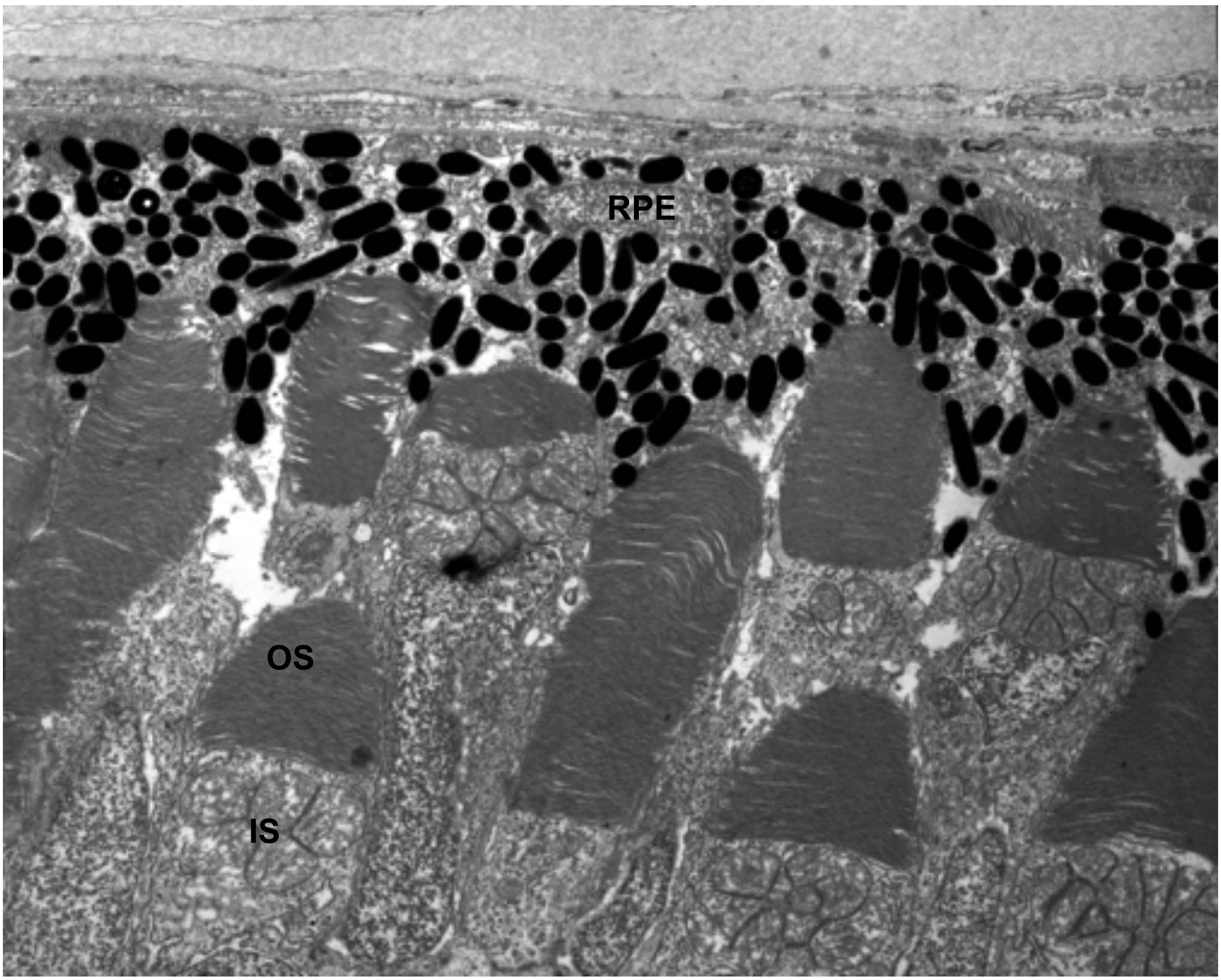
Fig. S3 Organization of outer segment disk membranes appears normal in 6dpf ush1cMO larvae. A Transmission electron micrograph showing photoreceptor outer segment ultrastructure in morphant larvae. RPE: Retinal Pigmented Epithelium; OS: Outer Segment; IS: Inner segment. Scale bar: 1μm.
Acknowledgments
This image is the copyrighted work of the attributed author or publisher, and
ZFIN has permission only to display this image to its users.
Additional permissions should be obtained from the applicable author or publisher of the image.
Full text @ Dis. Model. Mech.

