
|
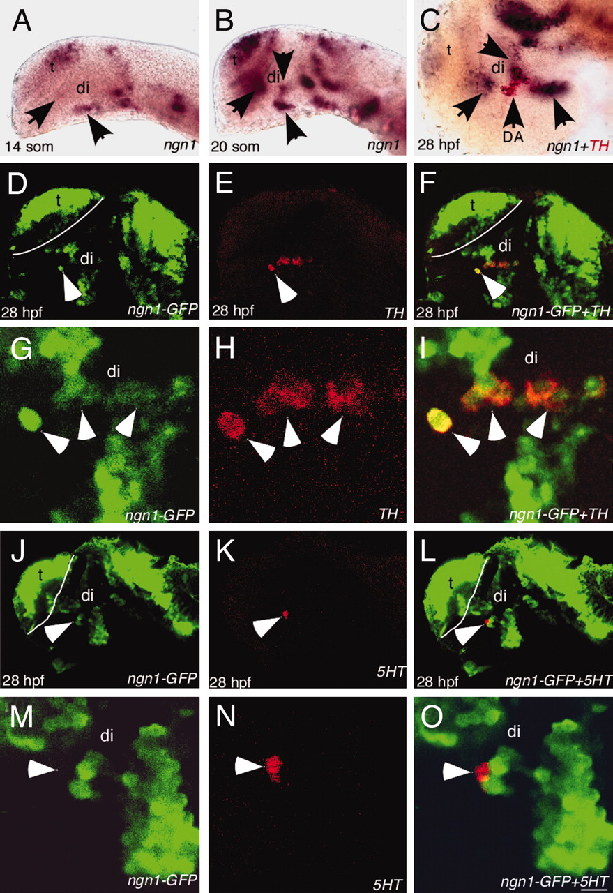
Fig. 1
ngn1 is expressed in DA progenitors. All images are lateral views of anterior brain regions. Anterior is to the left, and dorsal is up. (A and B) ngn1 expression at 14- and 20-somite stages, respectively (arrows point to several clusters in the ventral forebrain). (C) A 28-hpf embryo showing ngn1 expression (purple) in close proximity to TH+ DA neurons (red). (D?F) Confocal images of 28 hpf ngn1-GFP transgenic embryos immunostained with GFP antibody (green, D), TH antibody (red, E), and the merged image (F), showing that GFP is detected in TH+ DA neurons. (G?I) High-magnification views of D?F. (J?L) Confocal images of 28-hpf ngn1-GFP transgenic embryos immunostained with GFP antibody (green, J), 5HT antibody (red, K), and the merged image (L), showing that GFP is not detected in 5HT neurons. (M?O) High-magnification views of J?L. di, diencephalon, t, telencephalon. (Scale bars, 64 μm in A and B, 60 μm in C?F and J?L, and 3 μm in G?I and M?O.)