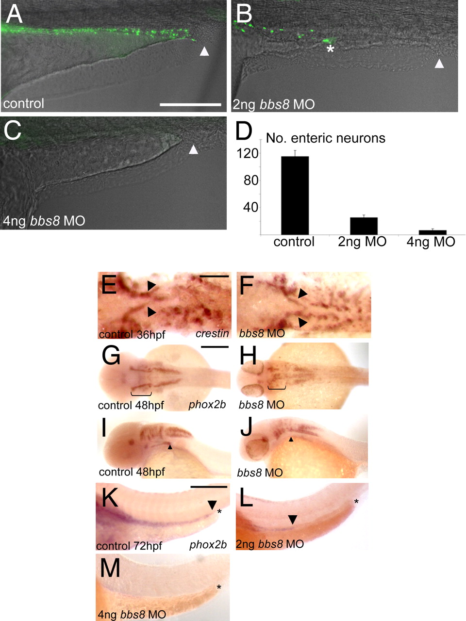
Fig. 5
Enteric nervous system development in bbs morphants. (A) Control embryos at 4 dpf showing enteric neurons populating the length of the gut from the anterior (left-hand side) to the anus (arrowhead). (B) With 2 ng of bbs8, MO migration of enteric neurons ceases midway along the gut (asterisk), and no neurons reach the anus (arrowhead). (C) Four nanograms of bbs8 MO causes complete absence of enteric neurons, resulting in gut motility defects (see Movie S4 and Movie S5). (D) Quantification of the reduction in number of enteric neurons with progressively higher doses of bbs8 MO. (E and F) NCCs exiting the vagal region visualized by crestin show no difference between control and morphant. (G and H) phox2b expression at 48 hpf shows a reduced number of NCCs in the branchial arches. (I and J) Lateral view of the embryos in G and H shows a failure of cells to leave the pharyngeal arches and enter the gut tube. (K) phox2b-positive enteric neurons populating the entire length of the gut all the way to the anus. The arrowhead shows the posteriormost extent of migration, and the asterisk represents the anus. (L and M) Injection of 2 ng of, and 4 ng of, bbs8 MO, respectively, causes neurons to migrate only halfway along the gut (2 ng) or fail to enter the gut at all (4 ng). [Scale bars: 500 μm (A?D), 100 μm (E and F), 200 μm (G?J), and 300 μm (K?M).]

