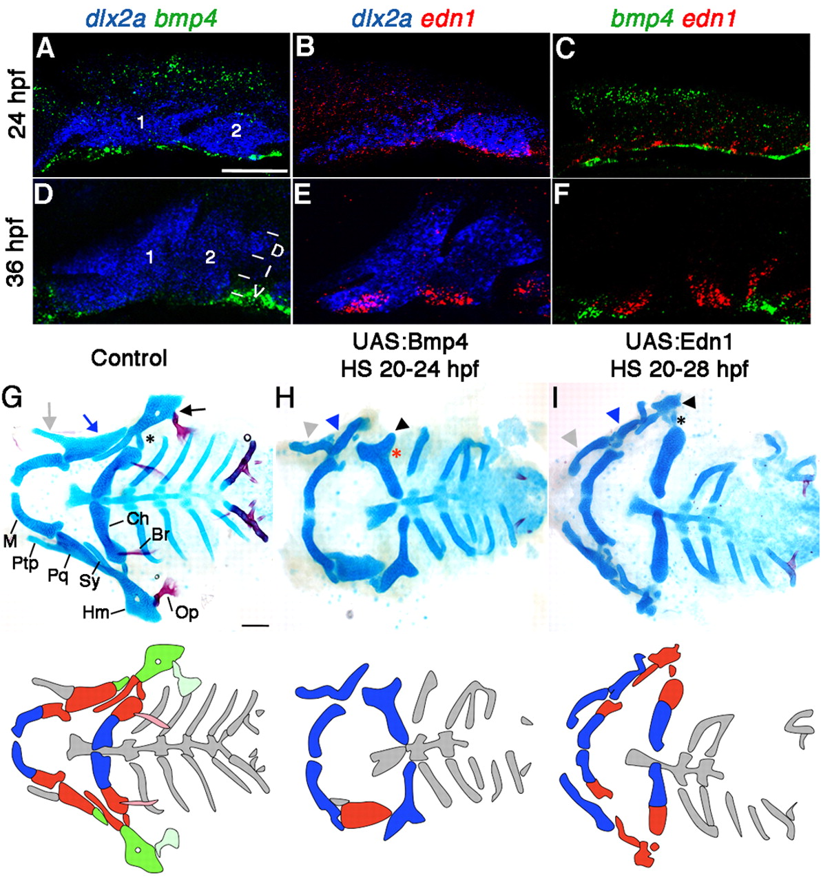
Fig. 1 Facial skeletal defects upon Bmp4 or Edn1 misexpression. (A-F) Confocal projections of in situ hybridization for dlx2a (blue), bmp4 (green) and edn1 (red) at 24 hpf (A-C) and 36 hpf (D-F) in wild type. Mandibular (1) and hyoid (2) arches are labeled, as well as dorsal (D), intermediate (I) and ventral (V) domains. (G-I) Ventral views (top) and schematics (below) of 5 dpf facial skeletons in control hsp70I:Gal4 (G) and hsp70I:Gal4; UAS:Bmp4 larvae subjected to a 4-hour heat-shock (H), and hsp70I:Gal4; UAS:Edn1 larvae subjected to an 8-hour heat-shock (I). Cartilage is blue and bone red. Schematics show dorsal (green), intermediate (red) and ventral (blue) regions with dermal bones lightly shaded. Hm (black arrow), Pq (blue arrow) and Ptp (grey arrow) were transformed (arrowheads) in UAS:Bmp4 and UAS:Edn1 larvae. In the intermediate second arch, the joint (asterisk) and symplectic bone were lost in UAS:Bmp4 but not UAS:Edn1 larvae. M, Meckel?s cartilage; Pq, palatoquadrate cartilage; Ptp, pterygoid process; Sy, symplectic cartilage; Hm, hyomandibular cartilage; Ch, ceratohyal cartilage; Op, opercular bone; Br, branchiostegal ray bone. Scale bars: 50 μm.
Image
Figure Caption
Figure Data
Acknowledgments
This image is the copyrighted work of the attributed author or publisher, and
ZFIN has permission only to display this image to its users.
Additional permissions should be obtained from the applicable author or publisher of the image.
Full text @ Development

