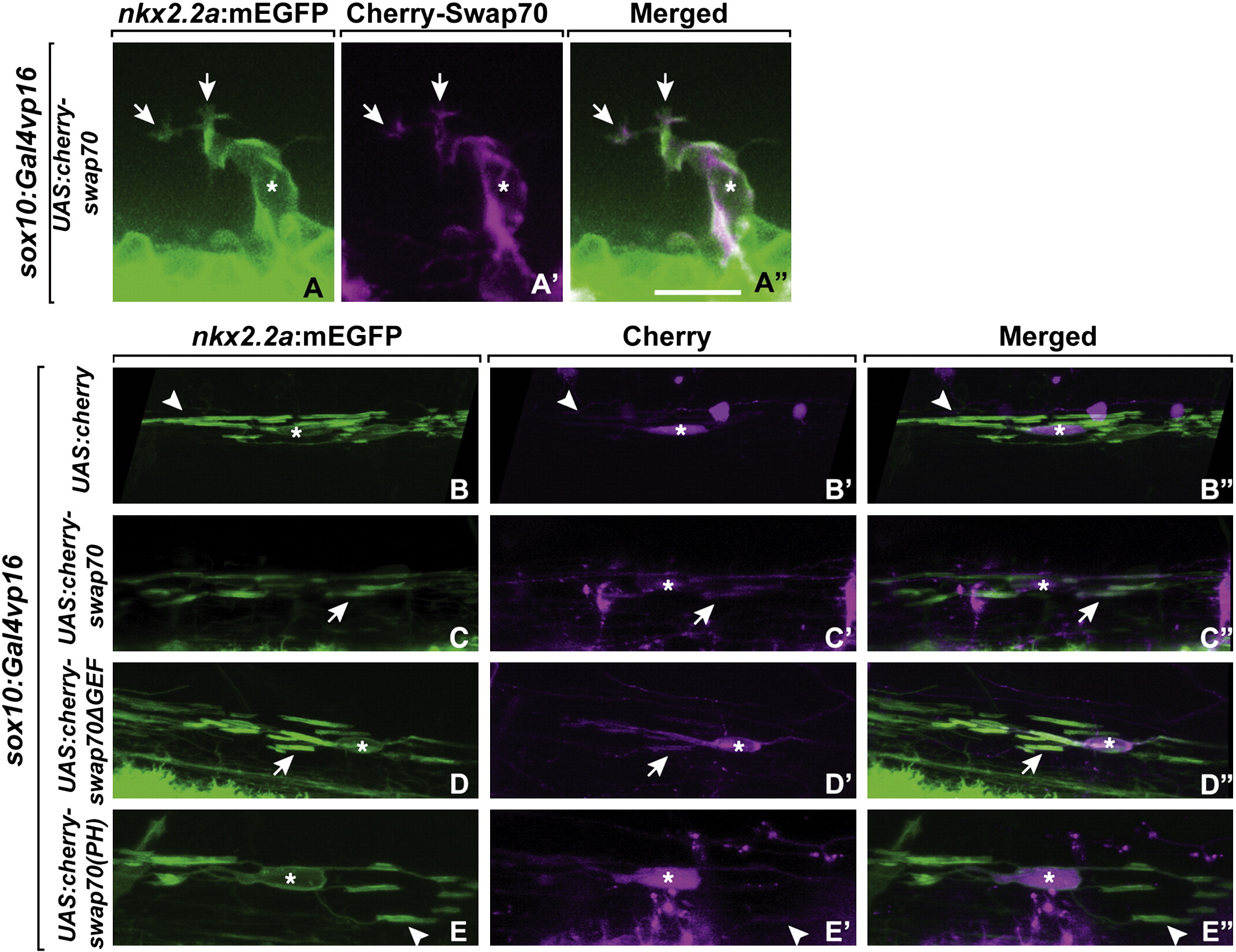
Fig. 4
Swap70 localization in migrating OPCs and myelinating oligodendrocytes. Cherry-Swap70 was transiently expressed in OPCs by co-injecting sox10:Gal4vp16 plasmid and the following plasmids: UAS:Cherry-Swap70 (A–A3, C–C3), UAS-Cherry (B–B3), UAS-Cherry-Swap70ΔGEF (D–D3), UAS-Cherry-Swap70(PH) (E–E3). (A–A3) Cherry-Swap70 was localized at the tips of membrane processes (arrows) and the cell membrane of migrating OPCs. (B–B3) In myelinating oligodendrocytes, Cherry localized to the cytoplasm (asterisks) but not to internode membrane (arrowheads). (C–C3) Cherry-Swap70 was localized to membrane (asterisks) and the axon-wrapping internodes (arrows). (D–D2) C-terminal deleted Swap70 fused to Cherry (Cherry-Swap70ΔGEF) was also localized in the membrane and axon wrapping internodes. (E–E3) Cherry-fused PH domain of Swap70 (Cherry-Swap70(PH)) was localized to the cytoplasmic and nuclear compartments (asterisks) but not internode membrane (arrowheads). Scale bar in A, 15 μm.
Reprinted from Molecular and cellular neurosciences, 48(3), Takada, N., and Appel, B., swap70 Promotes neural precursor cell cycle exit and oligodendrocyte formation, 225-35, Copyright (2011) with permission from Elsevier. Full text @ Mol. Cell Neurosci.

