
Fig. 2
|
Fig. 2
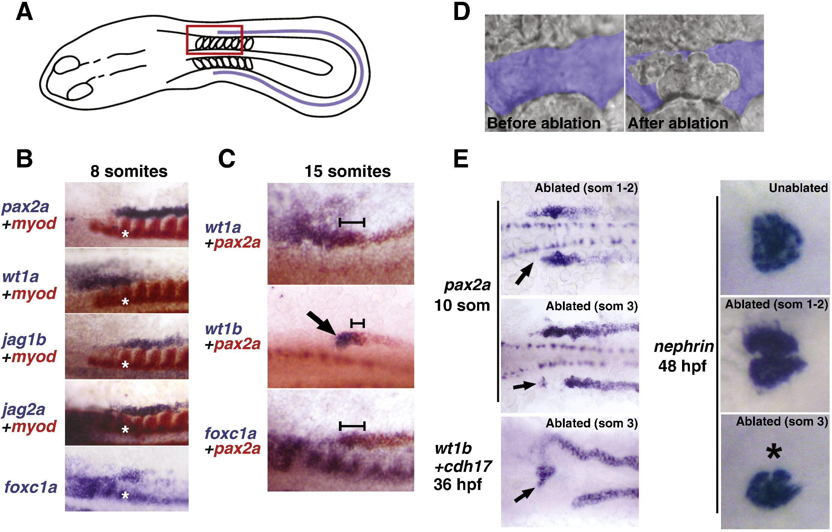
Podocytes arise from intermediate mesoderm adjacent to somite 3. A) Schematic of a flat-mounted 8-somite embryo showing the region of intermediate mesoderm (blue line) next to the somites (ovals) that was analyzed for expression of podocyte transcription factors or associated pathway components. Red box represents the regions shown in B) and C). B) In situ hybridization showing the expression patterns of pax2a, wt1a, foxc1a and the notch ligands jag1b and jag2a. Overlapping expression of all factors is seen in the region next to somite 3, marked by the asterisk. C) Micrographs showing overlapping expression of wt1a, wt1b and foxc1a with pax2a at the level of somite 3. Arrow points to overlapping expression of wt1b and pax2a in the anteriormost pax2a region (presumptive podocyte progenitors). D) DIC images of tissue adjacent to somite 3 analyzed by laser ablation. The top image highlights the region of intermediate mesoderm in blue prior to ablation. The image on the bottom shows the region after ablation, where the cells have lysed. E) In situ hybridization of ablated embryos at 10 somites (pax2a), 36 hpf (wt1b and cdh17) and 48 hpf (nephrin). Ablations were performed at the level of somites 1?2 or somite 3. Arrows point to remaining areas of pax2a and wt1b expression on the ablated side of 10 somite and 36 hpf embryos, respectively. The asterisk in the bottom-right panel marks the lack of nephrin expression in the ablated half of the embryo.
Reprinted from Developmental Biology, 358(2), O'Brien, L.L., Grimaldi, M., Kostun, Z., Wingert, R.A., Selleck, R., and Davidson, A.J., Wt1a, Foxc1a, and the Notch mediator Rbpj physically interact and regulate the formation of podocytes in zebrafish, 318-30, Copyright (2011) with permission from Elsevier. Full text @ Dev. Biol.