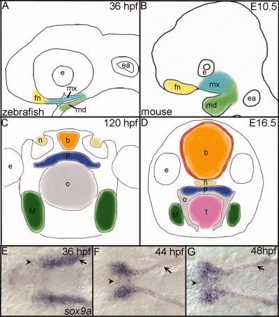
Fig. 1
Comparison of zebrafish and mouse palatogenesis with a detailed look at zebrafish palate formation. A,B: Outlines of embryos (lateral view), after CNCC condensation in the frontonasal (yellow), maxillary (turquoise), and mandibular (green) domains of zebrafish (A) and mouse (B). C,D: Drawings of cross-sections through heads of zebrafish (C) and mouse (D) after formation of the palate. Colors correspond to similar structures across both species. E,F,H: In situ hybridization of sox9a expression marking the palatal CNCC in zebrafish, ventral view with anterior to the left is shown, and mandibular crest have been removed to allow full view of the developing palate. E: At 36 hours postfertilization (hpf), sox9a expression marks the CNCC that line the maxillary (arrow) and the frontonasal (arrowhead) domains before their migration to the midline. F: Migration of frontonasal cells to the midline to begin the formation of the ethmoid plate (arrowhead) at 44 hpf and maxillary cells (arrow) begin rearrangements to form the trabeculae rods of the palate. G: By 48 hpf, most, if not all, frontonasal CNCC have migrated to the midline (arrowhead) and trabeculae rods are well formed in the palate (arrow). fn, frontonasal CNCC; mx, maxillary CNCC; md, mandibular CNCC; e, eye; ea, ear; b, brain; n, nasal opening; p, palate; o, oral opening; t, tongue; M, Meckel′s.

