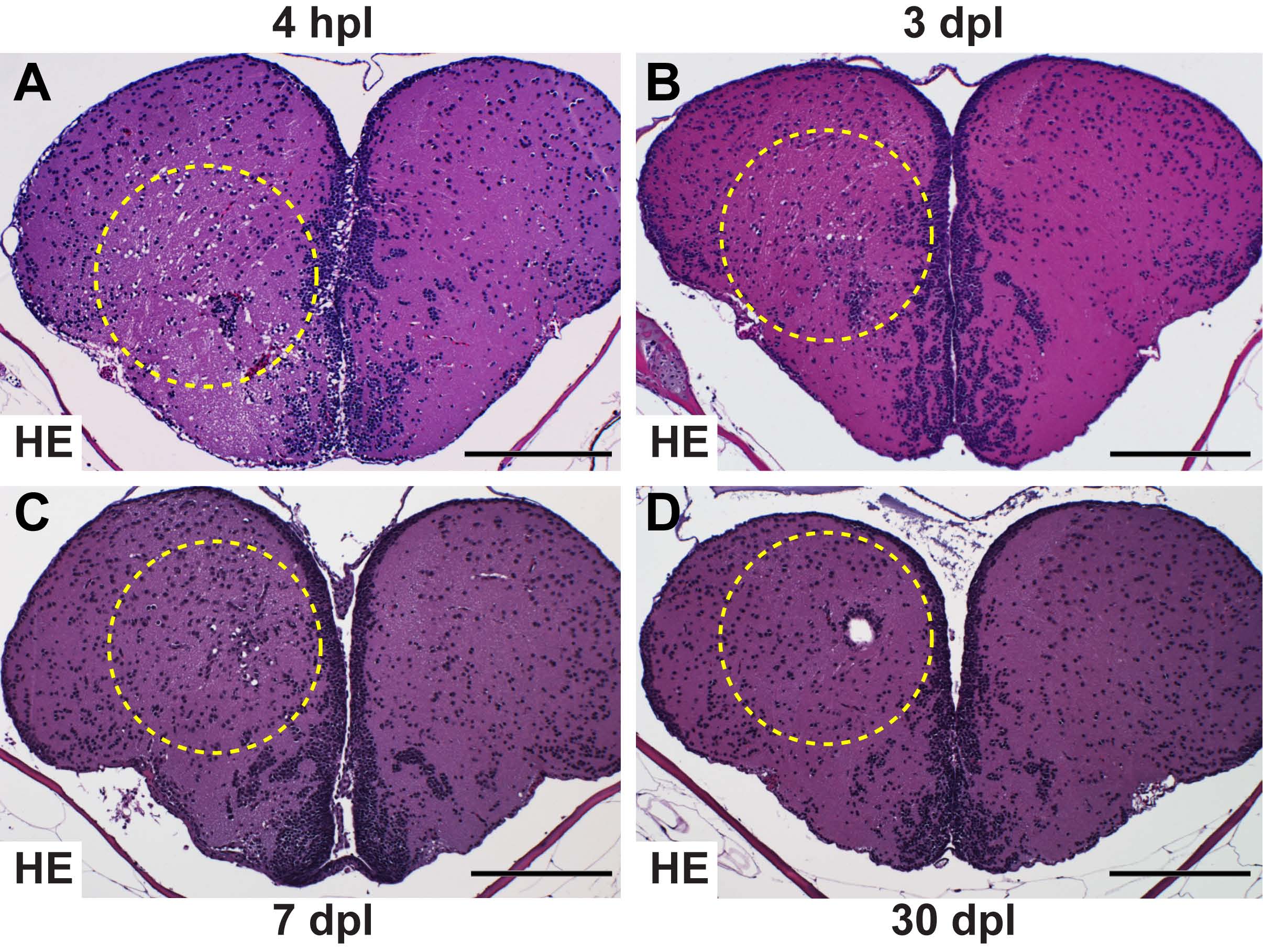
Fig. S1 Timecourse of status spongiosus and re-establishment of tissue integrity. (A) At 4 hpl the majority of the cross-sectional area of the lesioned hemisphere has the typical sieve-like structure known as status spongiosus. In addition, tissue outside the stab canal shows a spongious phenotype due to oedema. (B) By 3 dpl, the vacuolated area is dramatically reduced in size compared with at 4 hpl (A). The size of the vacuoles is strongly reduced compared with 1 dpl (Fig. 1B). (C) The oedema size and the degree of vacuolisation is further decreased at 7 dpl compared with 3 dpl. (D) The oedema size is further reduced at 30 dpl. Often a single small hole is seen. Scale bars: 200 μm. All panels show HE stains of 1 μm paraffin sections. Dashed outlines represent the lesion canal.
Image
Figure Caption
Acknowledgments
This image is the copyrighted work of the attributed author or publisher, and
ZFIN has permission only to display this image to its users.
Additional permissions should be obtained from the applicable author or publisher of the image.
Full text @ Development

