
|
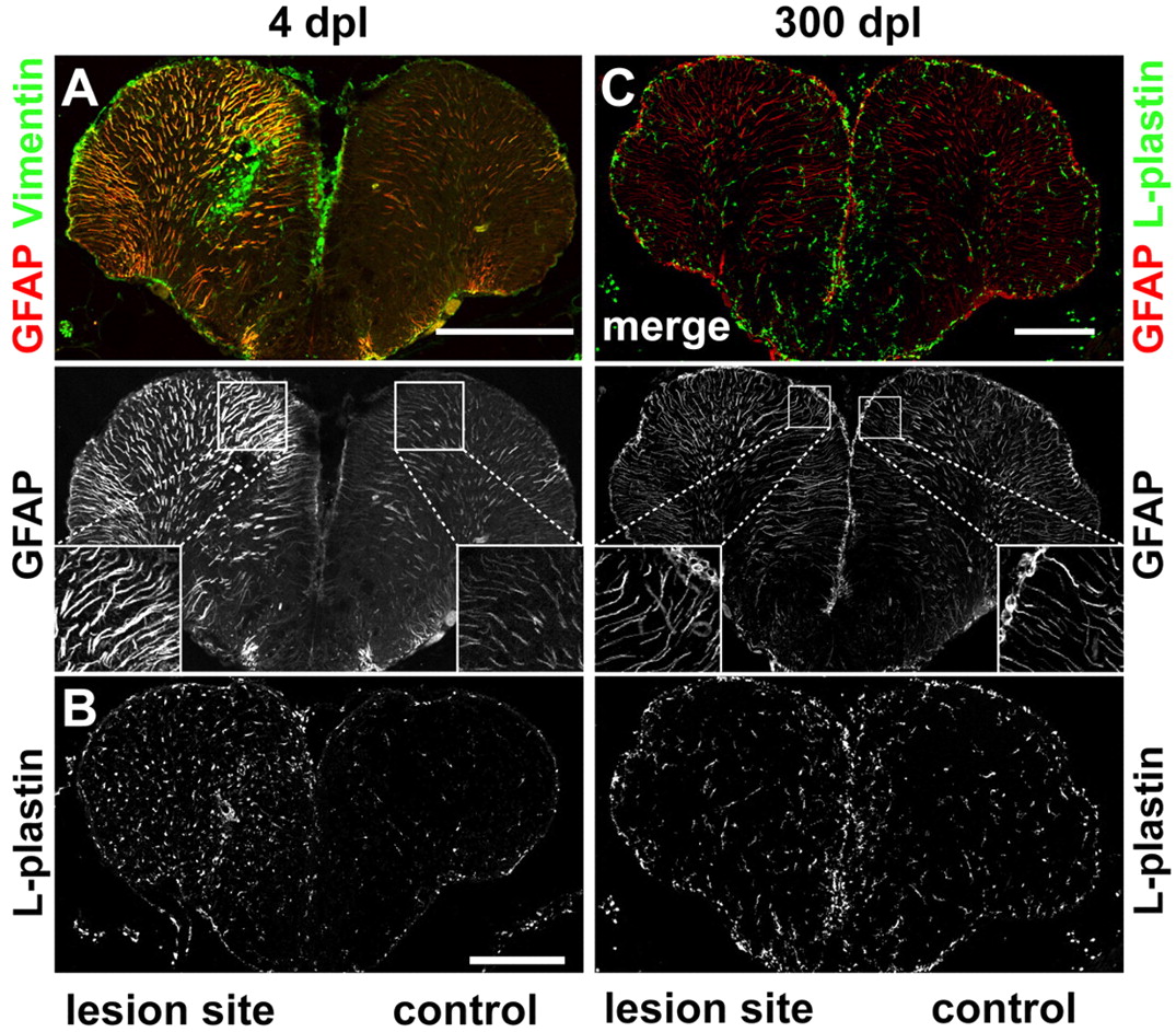
Fig. 2 Reactive gliosis, inflammatory response and scarring. (A) The glial markers GFAP (red) and vimentin (green) are strongly upregulated in the lesioned hemisphere early after injury (4 dpl). GFAP+ radial fibres in the lesioned hemisphere are thicker compared with the control hemisphere. (B) The pan-leukocyte marker L-plastin is strongly upregulated in the lesioned hemisphere at 4 dpl. (C) No difference in number and appearance of GFAP+ (red) radial processes is detected comparing lesioned and control hemispheres 300 dpl. Staining for L-plastin (green) shows no difference in the number and distribution of leukocytes between the lesioned and the control hemisphere. Scale bars: 200 μm.