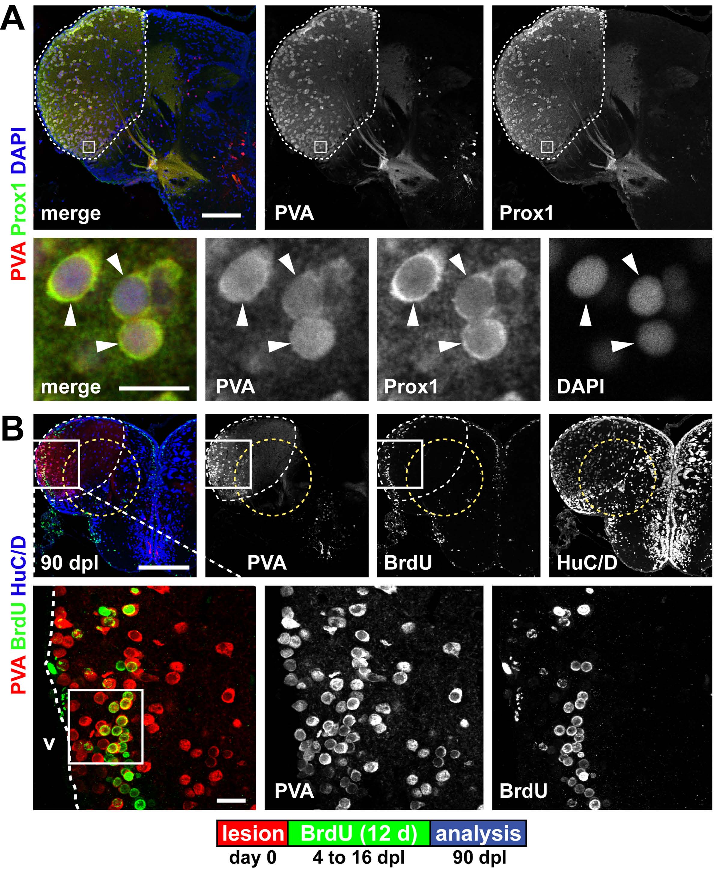
Fig. S9 Parvalbumin and Prox1 expression mark a territory within the lesion site in the dorsolateral telencephalon. (A) A territory in the dorsolateral telencephalon is characterized by the co-expression of the interneuron marker parvalbumin (PVA, red) and Prox1 (green) in a neuronal subpopulation (white dashed outline). Co-expression (arrowheads) is shown in a single confocal section. (B) The PVA/Prox1 territory (white dashed outline) is further characterized by weak expression of HuC/D (blue) and reaches into the lesion canal (yellow dashed circle). When BrdU is applied 4 to 16 dpl to label newborn cells, many periventricular PVA+ (red)/BrdU+ (green) are found at 90 dpl within the dorsolateral telencephalon, as shown in confocal max projections. This shows that the lesion does not alter the fate of newborn neurons: they acquire a spatially appropriate neuronal subtype. The area framed in the inset indicates the location of the single confocal section displayed in Fig. 5A. Scale bars: 200 μm; 50 μm in inset.
Image
Figure Caption
Acknowledgments
This image is the copyrighted work of the attributed author or publisher, and
ZFIN has permission only to display this image to its users.
Additional permissions should be obtained from the applicable author or publisher of the image.
Full text @ Development

