Image
Figure Caption
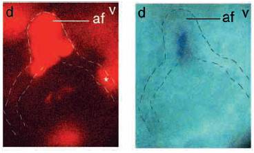
Fig. 8 Transplantation of wt cell into a dak- fin bud. Distal is towards the top, dorsal towards the left and ventral towards the right. Fluorescent image (left) shows biotin-labeled wt cell in the apical fold (af) and in the ventral non-ridge ectoderm (*). Bright-field image (right) shows expression of fgf8 in the apical fold. The outline of the bud and the epidermal basal lamina are indicated by the broken lines.
Acknowledgments
This image is the copyrighted work of the attributed author or publisher, and
ZFIN has permission only to display this image to its users.
Additional permissions should be obtained from the applicable author or publisher of the image.
Full text @ Development

