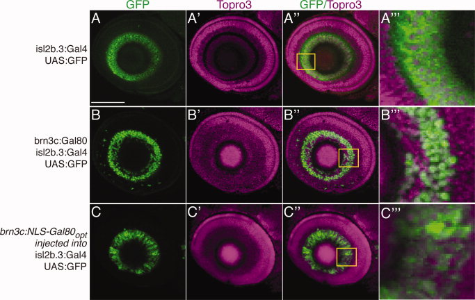
Fig. 5 Nuclear localization and codon optimization improves function of Gal80. Confocal maximum projections, lateral views, anterior to left, dorsal up, of eyes in Tg(isl2b.3:Gal4)zc65; Tg(UAS:GFP) 72 hours postfertilization (hpf) embryos. Immunostaining for green fluorescent protein (GFP), green; Topro3 nuclear stain, magenta. A?A′′ ′: Tg(isl2b.3:Gal4)zc65; Tg(UAS:GFP) transgenic embryos with no Gal80 show GFP expression in all retinal ganglion cells (RGCs). Inset and A′′ ′ shows high power magnification of single confocal slice. B?B′′ ′: Triple transgenic Tg(isl2b.3:Gal4)zc65; Tg(UAS:GFP); Tg(brn3c:Gal80) shows inhibition of Gal4-dependent GFP expression in approximately 30% of RGCs. Inset and B′′ ′ shows high power magnification of single confocal slice. C?C′′ ′: Transient injection with construct carrying ?improved? Gal80 into Tg(isl2b.3:Gal4)zc65; Tg(UAS:GFP) embryos demonstrates inhibition similar to that of stable lines carrying native Gal80. Improved Gal80 has nuclear localization signal (NLS) and is codon optimized (?opt?). Scale bar = 50 μm.
Image
Figure Caption
Acknowledgments
This image is the copyrighted work of the attributed author or publisher, and
ZFIN has permission only to display this image to its users.
Additional permissions should be obtained from the applicable author or publisher of the image.
Full text @ Dev. Dyn.

