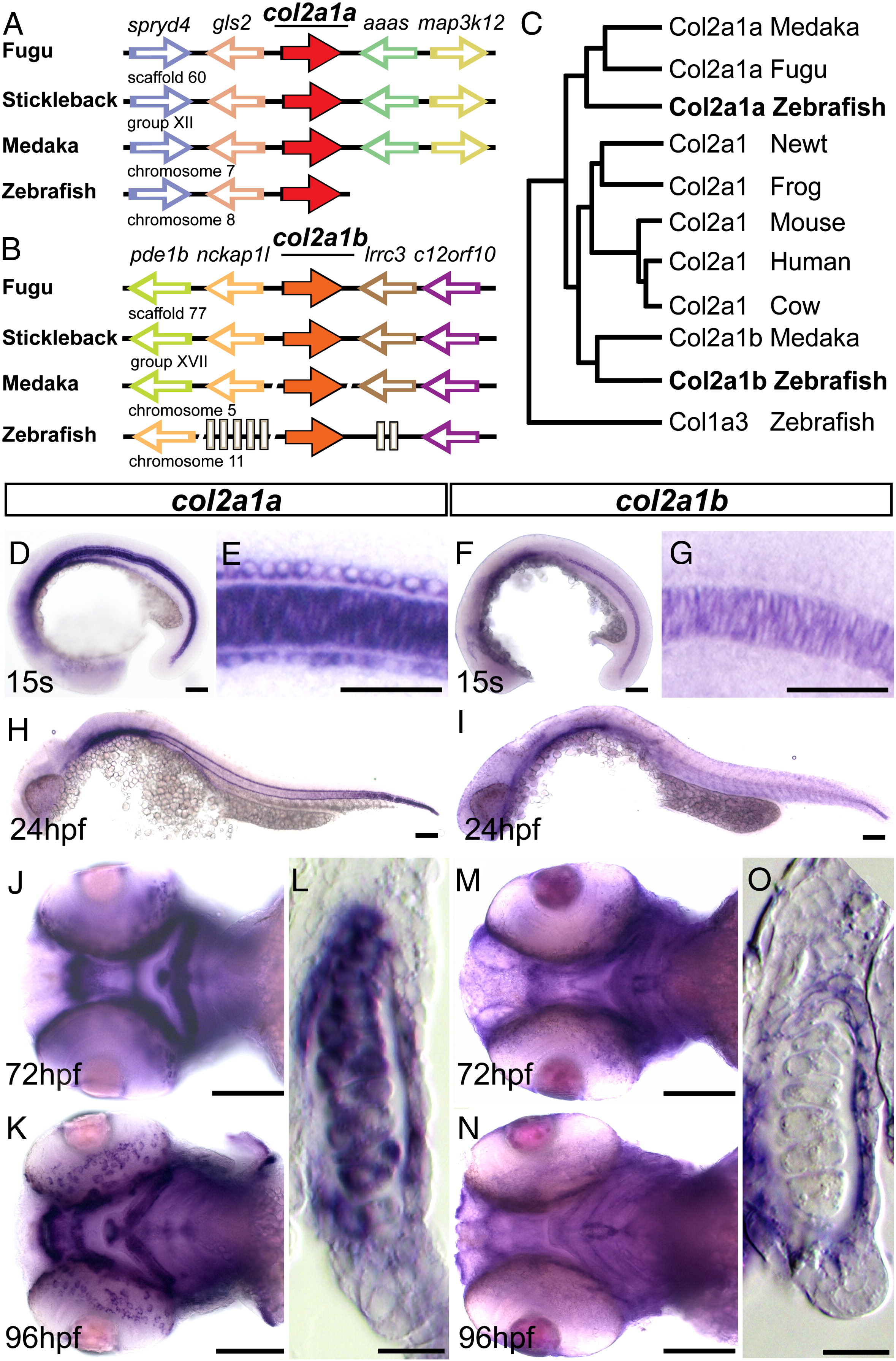
Fig. 1 Conservation and expression of zebrafish col2a1 homologues. A?B. Schematic of conserved genomic synteny around the col2a1a (A) and col2a1b (B) genes in fugu, stickleback, medaka, and zebrafish with gene orientation marked by the direction of the arrows. Location of the genomic region is marked below. The angled slices through line denote gaps in the published sequence and grey boxes demark genes not conserved between teleosts. C. Phylogenic tree of the protein sequence between vertebrate col2a1 homologues. D?O. Developmental expression patterns of col2a1a (D, E, H, J?L) and col2a1b (F, G, I, M?O) genes. D, F. Lateral view of a 15 somite stage embryo. E. Magnification of trunk region of the embryo shown on panel (D) with expression in the floor plate, notochord, and hypochord. G. Magnification of trunk of the embryo (F) showing only notochord expression. H, I. Lateral view of 24 hpf embryo. J, M. Ventral view of 72 hpf head. K, N Ventral view of 96 hpf head. L, O. Transverse section through ceratohyal cartilage and surrounding tissue of a 96 hpf embryo. Scale bars = 50 μm except for E, G, L and O = 25 μm.
Reprinted from Developmental Biology, 357(2), Dale, R.M., and Topczewski, J., Identification of an evolutionarily conserved regulatory element of the zebrafish col2a1a gene, 518-31, Copyright (2011) with permission from Elsevier. Full text @ Dev. Biol.

