Image
Figure Caption
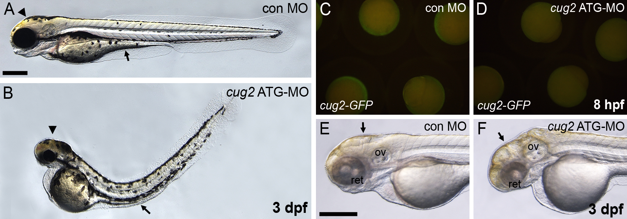
Fig. S1 Phenotypes of cug2 translation blocking morpholino (ATG-MO) in zebrafish embryos. A, B. cug2 MO-injected embryo (B) shows developmental defects including flat head, pinched midbrain-hindbrain boundary (arrowhead), thin yolk extension (arrow), and curved-up body. C, D. The translation blocking MO (ATG-MO) specifically inhibits the translation of cug2-GFP mRNA containing its targeting region. E, F. DIC image of PTU-treated cug2 morphant. The cug2 morphant shows retina degeneration and pinched brain structure (arrow) at 3 dpf. ov, otic vesicle; ret, retina. Scale bars = 200 μm.
Figure Data
Acknowledgments
This image is the copyrighted work of the attributed author or publisher, and
ZFIN has permission only to display this image to its users.
Additional permissions should be obtained from the applicable author or publisher of the image.
Full text @ BMC Dev. Biol.

