
|
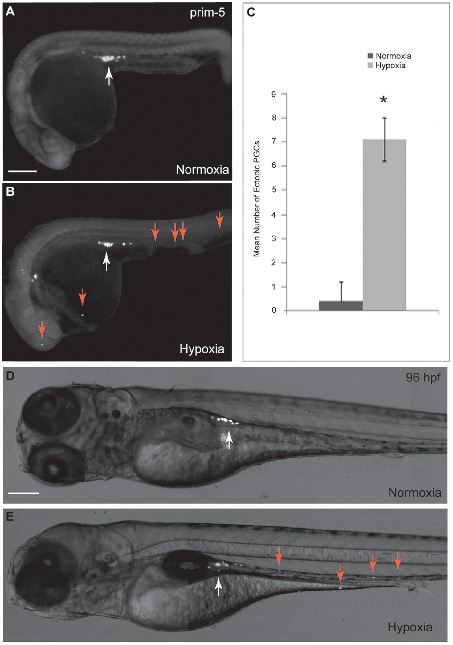
Fig. 1
Hypoxia affects PGC migration in zebrafish embryos.
Embryos were injected with GFP-nosl 32UTR mRNA as PGC marker. (A) PGCs in normoxic embryos migrated properly towards the genital ridge at prim-5 stage (white arrow). (B) PGC migration is affected by hypoxia as illustrated by presence of mis-migrated ectopic PGCs at prim-5 stage (red arrows). (C) Significantly greater number of mis-migrated ectopic PGCs was observed in hypoxic embryos as compared with normoxic embryos at prim-5 stage. (D) PGCs in normoxic embryos located at the genital ridge at 96 hpf. (E) Mis-migrated ectopic PGCs in yolk sac and in cranial region remained in the same position and failed to move towards the genital ridge at 96 hpf (red arrows). Scale bar: 200 μm. * denotes p<0.05.