Fig. 3
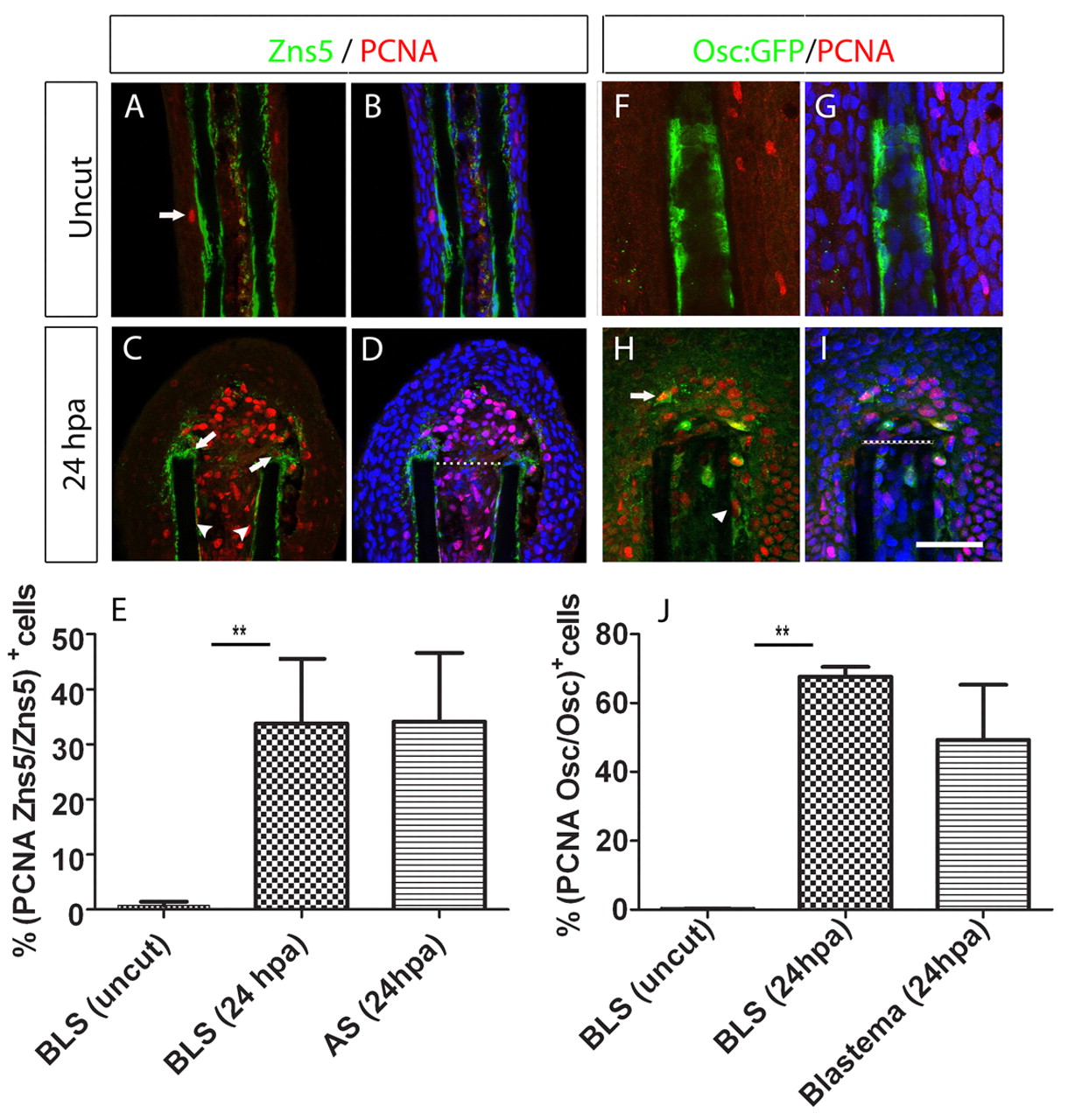
Proliferation of bone-lining cells increases upon amputation of the caudal fin. (A-D) Double immunohistochemistry with PCNA and Zns5 antibody in wild-type fins. (A,B) Uncut and (C,D) 24 hpa fins. (A,C) Zns5 (green) and PCNA (red). (B,D) Zns5 (green), PCNA (red) and DAPI (blue). The arrow in A indicates an proliferating epidermal cell. The arrowheads in C indicate proliferating BLSs and the arrows indicate aligning proliferating scleroblasts. (E) Percentage of the total Zns5-positive cells that were also positive for PCNA in the BLS and AS populations, calculated for the first segment below and above the amputation plane at 24 hpa, and in the equivalent region of uncut fins. There was a significant increase in double-positive BLSs in the 24 hpa samples compared with uncut samples (Student′s t-test, P<0.05; n=5 for each condition). (F-I) Double immunohistochemistry with GFP and PCNA antibody in Tg(osteocalcin:GFP) fins. (F,G) Uncut fin. (H,I) 24 hpa fin. (F,H) osteocalcin:GFP (green) and PCNA (red). (G,I) osteocalcin:GFP (green), PCNA (red) and DAPI (blue). In H, the arrow indicates a proliferating osteocalcin-expressing cell within the blastema; the arrowhead indicates a proliferating osteocalcin-expressing BLS. (J) Percentage of cells double-positive for osteocalcin:GFP and PCNA over the total osteocalcin:GFP-positive cells, in the BLS and blastema populations, calculated for the first segment below and above the amputation plane at 24 hpa, and in the equivalent region of uncut fins. There was a significant increase in double-positive BLSs in the 24 hpa samples compared with uncut samples (Student′s t-test, P<0.05; n=5-8 for each condition). Scale bar: 100 μm in A,B,F-I and 75 μm in C,D. Dashed lines indicate amputation plane.

