Fig. S6
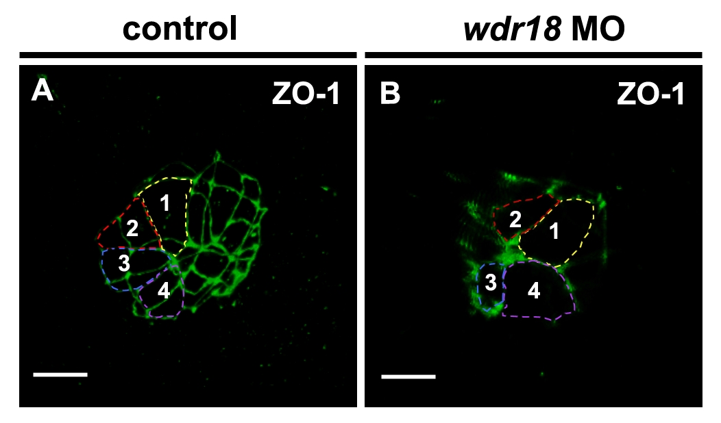
Knockdown of wdr18 did not change the size of cells composing Kupffer′s vesicle. 3D reconstruction confocal images of the KV in control and wdr18 morphant embryos revealed by immuno-staining with ZO-1 antibody at 6-somite stage. For the convenience of outlining the cell shape, the 3D images were rotated to the proper position until the boundaries of the marked cells was clearly seen. (A) In a control embryo, the cell connection was obvious and we outlined four cells with different colors and numbered them from 1 to 4. (B) In a wdr18 morphant, we similarly outlined and numbered four cells for comparison. The size of the marked cells showed no obvious difference comparing with that in control embryos. Scale bars: 15 μm.

