Fig. S3
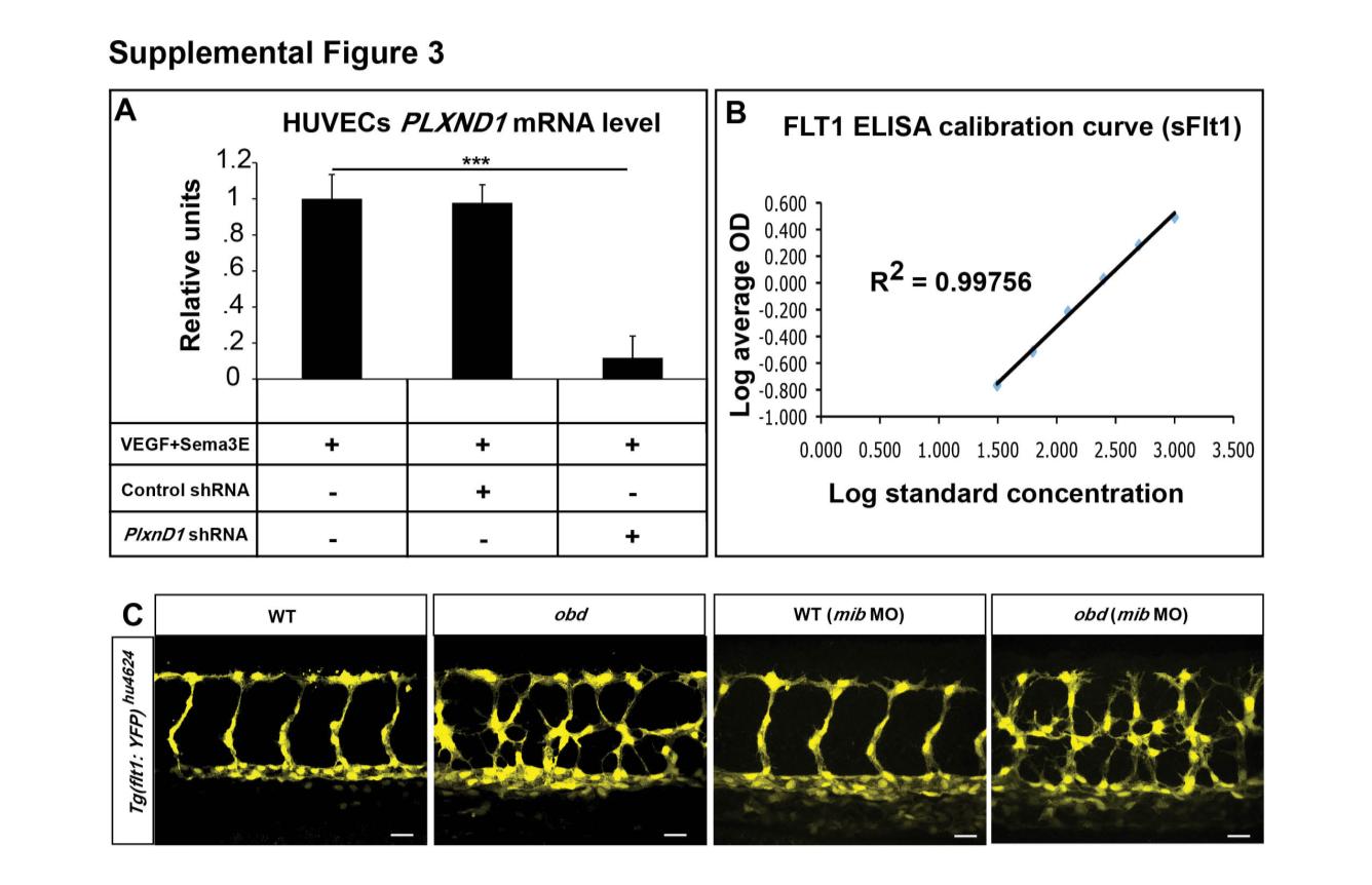
Related to Figure 3. PLXND1 mRNA Levels in HUVECs under Different Conditions, Experimental Controls for Flt1 ELISA and Expression of the flt1 Transcriptional Reporter Tg(flt1:YFP)hu4624 in WT and obd
(A) Validation of shRNA-mediated PLXND1 knockdown. qPCR measurements of PLXND1 mRNA abundance in HUVECs treated with VEGF and Sema3E and infected with the indicated control or PLXND1-targeting shRNA lentiviral particles. ***p < 0.001. Error bars, s.e.m. (B) Standard calibration curve generated for the ELISA-based quantification of Flt1 made by HUVECs based on average optical density (OD) at various concentrations. Coefficient of determination, R2 = 0.99756. Error bars, s.e.m. (C) Expression of the Tg(flt1:YFP)hu4624 reporter in WT and obd with or without 10 ng of mib morpholino (mib MO). n = 10 for WT, n = 12 for obd, n = 11 for WT (mib MO) and n = 9 for obd (mib MO).
Reprinted from Developmental Cell, 21(2), Zygmunt, T., Gay, C.M., Blondelle, J., Singh, M.K., Flaherty, K.M., Means, P.C., Herwig, L., Krudewig, A., Belting, H.G., Affolter, M., Epstein, J.A., and Torres-Vazquez, J., Semaphorin-PlexinD1 Signaling Limits Angiogenic Potential via the VEGF Decoy Receptor sFlt1, 301-314, Copyright (2011) with permission from Elsevier. Full text @ Dev. Cell

