Fig. 3
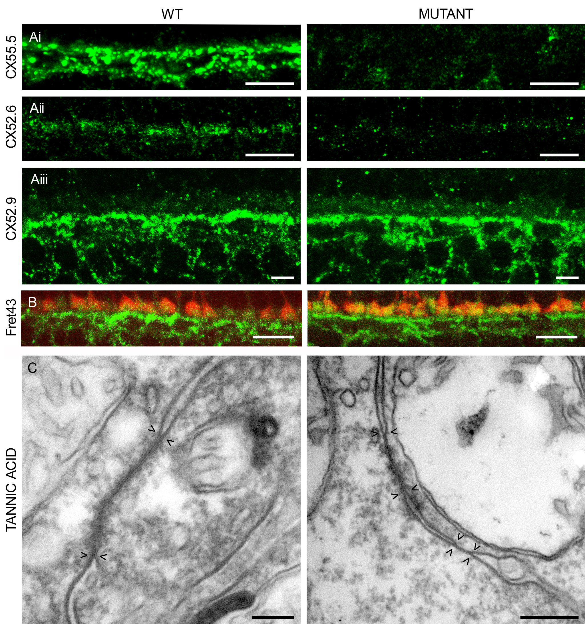
Localization and expression of connexins expressed by horizontal cells in wild-type and mutant zebrafish.
(A) Immunocytochemical staining with antibodies against Cx55.5 (i), Cx52.6 (ii), and Cx52.9 (iii). Cx55.5-IR and Cx52.6-IR are absent in the mutant, whereas Cx52.9-IR remains present or even becomes stronger. Scale bar = 10 μm. (B) Double labeling of Cx52.9 (green) and FRet43 (red), a label for double cones. The Cx52.9-IR and FRet43-IR are closely associated, indicating that Cx52.9-IR is present in the dendrites of horizontal cells invaginating the cone synaptic terminal in both wild-type and mutant. Scale bar = 10 μm. (C) Tannic acid staining of gap-junctions in wild-type and mutant retinas. Pairs of arrow heads indicate the extent of the gap-junction. Often the gap-junctions in the mutant seem to be ?split? as is illustrated in this figure. On average, gap-junctions are smaller in the mutant retinas. Scale bar = 0.5 μm.

