Fig. S7a
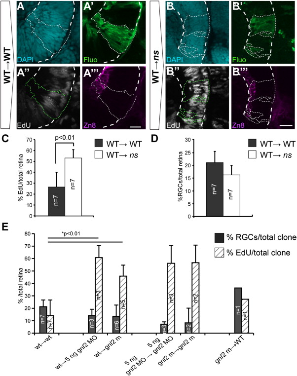
Gnl2 and NS act non-cell autonomously in retinal cell cycle exit and neuronal differentiation. (A and B) Single-plane confocal images of clones of transplanted cells showing DAPI-counterstained nuclei (cyan, top left), streptavidin-Alexa488 (green, top right), EdU (white, bottom left) and zn8-stained RGCs (magenta, bottom right). Clones of transplanted cells are outlined by dotted lines (white or green). The retina is outlined with dashed white lines. Shown are clones transplanted from WT donor to WT host (A) and WT donor to ns mutant host (B). (C and D) Quantification of the percentage of EdU+ (C) and zn8+ RGCs (D) within transplanted clones for WT→WT and WT→ns mutant. Statistics: Two-tailed Student′s t-test, p-values indicated above the bars (*p < 0.01, **p < 0.02). Error bars represent standard deviation. (E) Quantification of the percentage of EdU+ (striped bars) and zn8+ RGCs (black bars) within transplanted clones for additional experimental groups regarding gnl2 in addition to the data shown in Fig. 7F (with the exception of WT→WT and WT→gnl2 mutant which are shown in both Fig. 7F and here).
Reprinted from Developmental Biology, 355(2), Paridaen, J.T., Janson, E., Utami, K.H., Pereboom, T.C., Essers, P.B., van Rooijen, C., Zivkovic, D., and Macinnes, A.W., The nucleolar GTP-binding proteins Gnl2 and nucleostemin are required for retinal neurogenesis in developing zebrafish, 286-301, Copyright (2011) with permission from Elsevier. Full text @ Dev. Biol.

