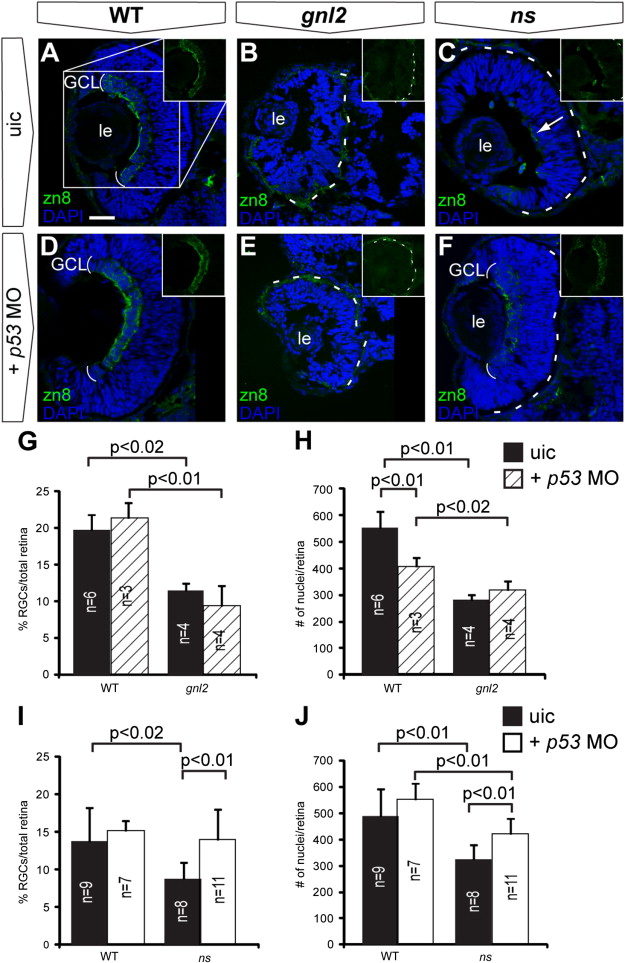
Fig. S6
Stabilization of p53 affects early retinal differentiation in ns but not gnl2 mutants. Confocal images of immunolabelings to the RGC marker zn8 (green) with DAPI nuclear counterstaining (blue) on coronal sections of uninjected control embryos (A?C) as compared to p53 MO-injected embryos (D?F). Injection of p53 MO did not affect RGC differentiation in WT (A and D), nor did it improve RGC differentiation in gnl2 mutant (B and E) embryos at 48 hpf. In contrast, in the ns mutant, injection of p53 MO resulted in the development of a distinct RGC layer at 48 hpf (F), whereas the uninjected ns mutants had a poorly formed RGC layer (arrow in C). Insets show magnification of single zn8 fluorescence (as indicated in box in A). The dashed lines indicate the outline of the retina. (G) Quantifications of the proportion of zn8+ cells in the total retina in WT and gnl2 mutants with or without injection of 1 ng of p53 MO. Upon p53 MO injection, the percentage of RGCs is unchanged in WT and gnl2 mutants at 48 hpf. (H) Quantification of the total number of retinal cells shows that p53 MO does not improve the reduction in cell counts in the gnl2 mutant retina. (I and J) Quantifications of the proportion of zn8+ cells (I) and total number of cells (J) in WT and ns mutant retina at 48 hpf with or without injection of 1 ng of p53 MO. Upon p53 MO injection, the percentage of RGCs is significantly increased in ns mutants (I). In addition, p53 MO injection significantly increases the total number of cells found in the retina (J), although it is still reduced as compared to WT embryos. Statistics: Student′s t-test, p-values and experimental numbers shown in graph. Error bars represent SD. GCL, ganglion cell layer; le, lens; uic, uninjected control. Scalebar 25 μm.
Reprinted from Developmental Biology, 355(2), Paridaen, J.T., Janson, E., Utami, K.H., Pereboom, T.C., Essers, P.B., van Rooijen, C., Zivkovic, D., and Macinnes, A.W., The nucleolar GTP-binding proteins Gnl2 and nucleostemin are required for retinal neurogenesis in developing zebrafish, 286-301, Copyright (2011) with permission from Elsevier. Full text @ Dev. Biol.

