
|
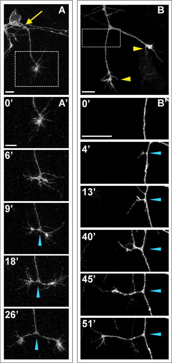
Fig. 3 Peripheral axons branch by bifurcation and interstitial branching. Individual RB neurons labeled by transient mosaic expression of GFP-CAAX in wild-type embryos. Lateral views, anterior is left. Images are confocal projections. (A) An unbranched peripheral axon formed at a site posterior to the RB cell body (yellow arrow). Box shows area imaged in (A′). (A′) Time-lapse of peripheral axon bifurcation (blue arrowheads indicate branch point). (B) A branched peripheral axon with growth cones extending ventrally (yellow arrowheads). Box shows area imaged in (B′). (B′) Time-lapse of interstitial branch formation (blue arrowheads indicate branch point). Time is displayed in minutes. Scale bars = 10 μm.