Fig. 5
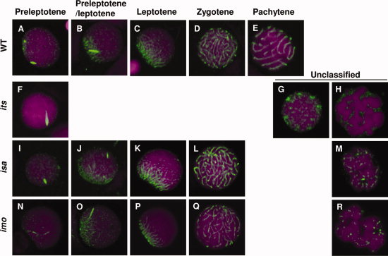
Immunocytochemical analyses of Sycp3 in spermatocytes from the its, isa, and imo mutants. Spermatocytes from wild-type (A?E), its (F?H), isa (I?M), and imo (N?R) were stained with anti-Sycp3 antibodies (green) and counterstained with TOPRO-3 (magenta). Each developmental stage of the spermatocytes was identified according to their synaptonemal complex (SC) structures. A?E: Localization of Sycp3 in wild-type spermatocytes at the preleptotene (A), leptotene?zygotene transition (B), leptotene (C), zygotene (D), and pachytene (E) stages. F?H: Localization of Sycp3 in its spermatocytes. The most progressed its spermatocytes were similar to wild-type spermatocytes at the preleptotene stage (F). Two types of abnormality were encountered; an accumulation of dotted SCs throughout the nucleus (G) and highly condensed chromosomes (H). I?M: Localization of Sycp3 in isa spermatocytes. L: The most progressed isa spermatocytes were similar to those of the wild-type zygotene stage. M: The most frequently encountered abnormal cells were associated with short fragments of the SC throughout the nucleus. N?R: Localization of Sycp3 in imo spermatocytes. Q: The most progressed imo spermatocytes were similar to wild-type zygotene stage spermatocytes. R: The most frequently encountered abnormal cells were associated with short fragments of the SC and slightly condensed chromosomes throughout the nucleus.

