
|
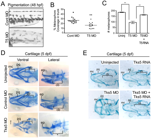
Fig. 2
Decreased Tks5 expression results in neural crest-derived defects.
(A?B) Melanophores within the trunk region above the yolk sac extension in control MO-injected and Tks5 MO-injected embryos were qualitatively (A) and quantitatively (B) analyzed. n = 15 embryos and SEM is shown by bar. p values obtained from Student′s t-test. ** denotes p<0.01. (C) Melanophores present in the dorsal, ventral, and lateral pigment lines were quantified to determine degree of murine Tks5 rescue of the decreased pigmentation seen in morphants. Mean values (n = 3) and SEM are shown in graph. p values obtained from Student′s t-test. ** denotes p<0.01. (D) Alcian blue staining was performed on indicated embryos to identify craniofacial structures (Meckel′s cartilage (mc), palatoquadrate (pq), ceratobranchials (ch), ethmoid plate (ep)). (*) denotes missing structures. (E) Alcian blue staining was performed on indicated embryos to determine if murine Tks5 could rescue craniofacial defects seen in morphants. Structures were identified as in (D). (*) denotes missing structures.