Fig. S3
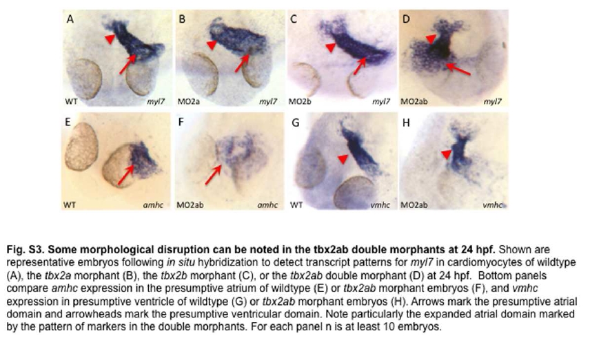
Some morphological disruption can be noted in the tbx2ab double morphants at 24hpf. Shown are representative embryos following in situ hybridization to detect transcript patterns for myl7 in cardiomyocytes of wildtype (A), the tbx2a morphant (B), the tbx2b morphant (C), or the tbx2ab double morphant (D) at 24 hpf. Bottom panels compare amhc expression in the presumptive atrium of wildtype (E) or tbx2ab morphant embryos (F), and vmhc expression in presumptive ventricle of wildtype (G) or tbx2ab morphant embryos (H). Arrows mark the presumptive atrial domain and arrowheads mark the presumptive ventricular domain. Note particularly the expanded atrial domain marked by the pattern of markers in the double morphants. For each panel n is at least 10 embryos.

