
|
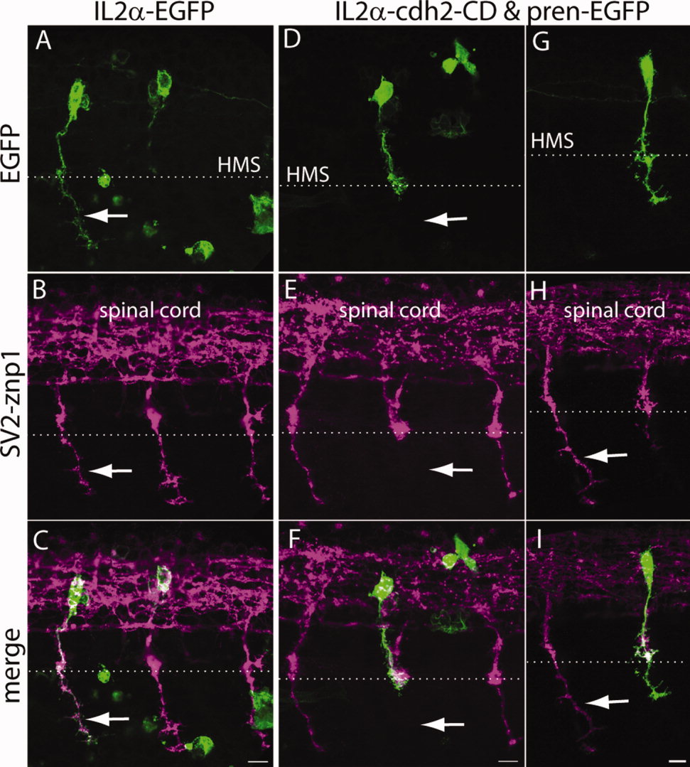
Fig. 9
Expression of N-cadherin dominant-interfering cytoplasmic domain perturbs CaP motor axon growth at the horizontal myoseptum. A–I: Tg(mnx1:Gal4-VP16) embryos were injected at the one-cell stage with IL2α-EGFP (A–C) or with IL2α-cdh2-CD and pren-EGFP (D–I) plasmids. Embryos were fixed at 24 hpf, immunostained with SV2 and znp1 and observed under confocal microscopy. A–C: Arrow points to the CaP axon extending ventral to the horizontal myoseptum (HMS), indicated with a dashed line. The IL2α-EGFP labeled axon has a normal morphology compared with the untransfected neighboring axons labeled with SV2 and znp1. D–F: Motor axon expressing IL2α-cdh2-CD grew through the common pathway but stalled at the horizontal myoseptum. The arrow points to the absence of SV2- and znp1-labeled CaP axon ventral to the choice point, whereas neighboring untransfected axons grew normally. G–I: CaP motor neuron expressing IL2α-cdh2-CD and pren-EGFP in which the axon extended ventrally to the horizontal myoseptum but showed a shorter migration distance compared with an untransfected SV2- and znp1-labeled axon (arrow). Dorsal is to the top and rostral is to the left. Scale bars = 10 μm in C (applies to A–C); 10 μm in F (applies to D–F); 10 μm in I (applies to G–I). [Color figure can be viewed in the online issue, which is available at wileyonlinelibrary.com.]