
|
Fig. 5
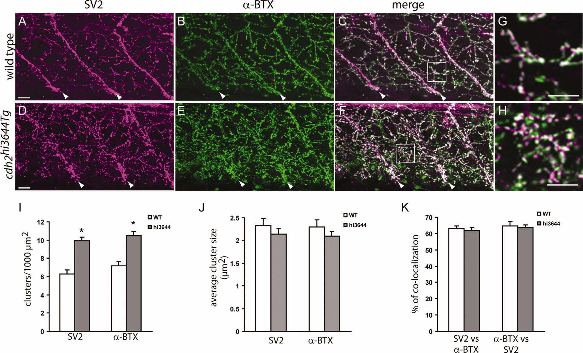
N-cadherin cdh2hi3644Tg mutants form higher numbers of neuromuscular junctions. Confocal images obtained from 120 hpf wild-type (A?C) and cdh2hi3644Tg (D?F) larvae doubly labeled with SV2 antibodies (A,D) and anti-mouse IgG Cy3-conjugated secondary antibody and with α-bungarotoxin (α-BTX) conjugated to Alexa 488 (B,E). Approximately 30-μm-thick stacks of confocal images obtained at 1-μm intervals were projected to a single plane. C,F: Merged images from A,B and D,E, respectively. Arrowheads point to the somitic myoseptum. G,H: High-magnification images of the boxed areas in C and F, respectively. I?K: Analysis of the number (I) and size (J) of SV2 and α-BTX clusters and of the percentage of colocalization between pre- and postsynaptic markers (K). Three confocal sections at the central region of the myotome were rendered to a single plane and used for quantification of the SV2 and α-BTX clusters. Open bars, wild type (WT; n = 16); gray bars, cdh2hi3644Tg (hi3644; n = 27; n, number of somitic hemisegments analyzed). Asterisks in I indicate a Student′s t-test P value < 0.005. Rostral is to the left and dorsal is to the top. Scale bars = 10 μm in A (applies to A?C); 10 μm in D (applies to D?F); 10 μm in G,H.