
IMAGE
Fig. 1
Image
|
Figure Caption
Fig. 1
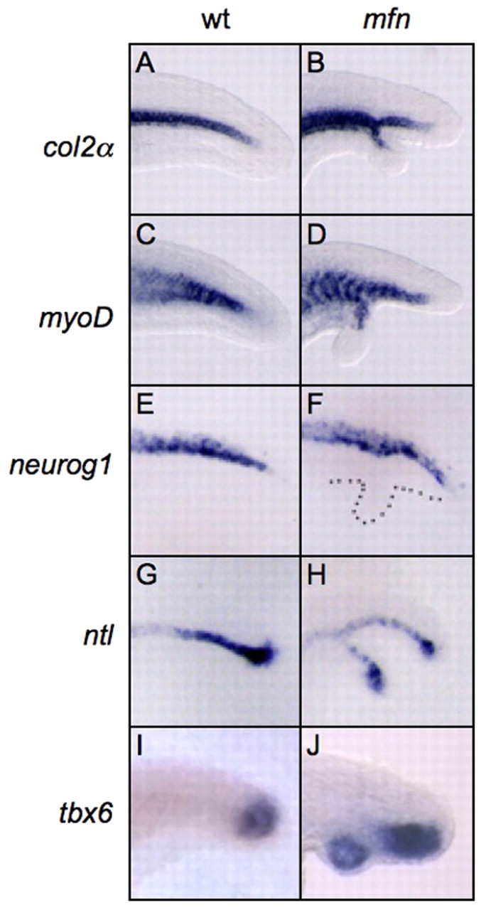
Mesoderm tissues are mis-specified in the secondary tails of mfn. (A-J) Lateral view of expression of col2a (col2α; A,B), myoD (C,D), neurog1 (E,F), ntl (G,H) and tbx6 (I,J) in the posterior tail of wild-type and mfn zebrafish embryos. At 26 hpf, col2a (B), myoD (D) and ntl (H) are ectopically expressed in the secondary tail, but neurog1 is not (F). At 24 hpf, tbx6 (J) is also ectopically expressed in the secondary tail. Embryos in all images are mounted with anterior to the left. Dotted line in F indicates secondary tail.
Figure Data
Acknowledgments
This image is the copyrighted work of the attributed author or publisher, and
ZFIN has permission only to display this image to its users.
Additional permissions should be obtained from the applicable author or publisher of the image.
Full text @ Development