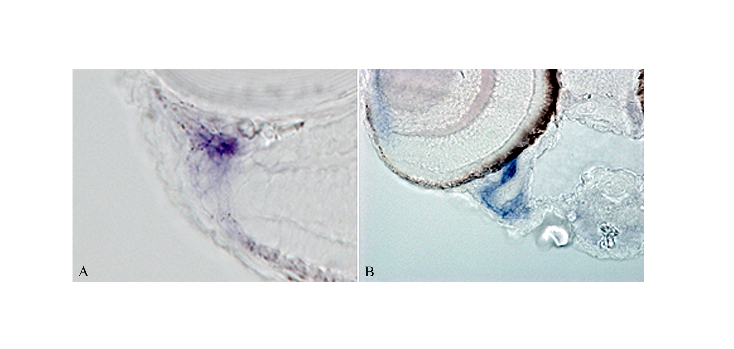
Fig. S1
mab21l2 is expressed in the ventral iridocorneal canal. A: At 5 dpf, cells expressing mab21l2 localise to the ventral aspect of the eye forming a circumferential structure, distinguished as a section of a channel. Expression within this structure is also detected in posterior serial sections, where it remains at the ventral aspect of the eye contiguous to the iridocorneal angle. B: These sections show a group of cells with a vessel-like shape that can be seen as an ipsilateral section of a tubular structure originating at the iridocorneal angle. The tubular organisation of the cells resembles the ventral canalicular network at the ventral iridocorneal angle, considered homologous to Schlemm′s canal in mammals.

