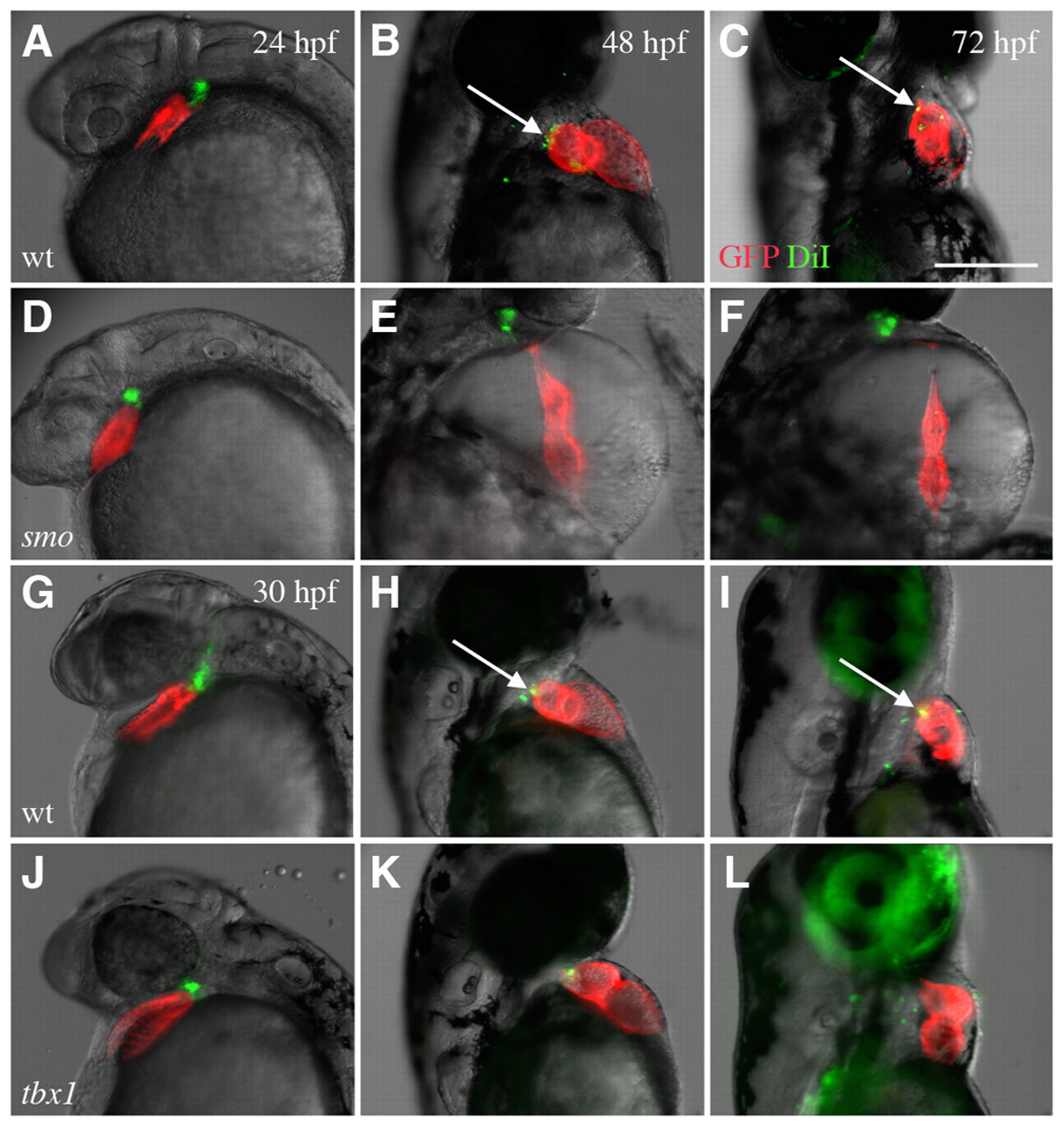
Fig. 9 Labeled cells in the pericardial wall are not incorporated into the developing heart in smo and tbx1 mutants. (A-C,G-I) Cells labeled with DiI (pseudocolored green) in the branchial region are incorporated into the myocardium (pseudocolored red) in wild-type zebrafish hearts after labeling at 24 hpf (A-C) and 30 hpf (G-I). DiI colocalized with the myocardium in wild-type fish at 48 and 72 hpf (arrows). (D-F,J-L) This incorporation of cells does not occur in most smo (D-F) or tbx1 (J-L) mutants. The green cells that appear to overlap with red cardiac cells (K) do not overlap with the myocardium 24 hours later (L). All embryos expressed cmlc2-GFP. Scale bar: 25 μm.
Image
Figure Caption
Acknowledgments
This image is the copyrighted work of the attributed author or publisher, and
ZFIN has permission only to display this image to its users.
Additional permissions should be obtained from the applicable author or publisher of the image.
Full text @ Development

