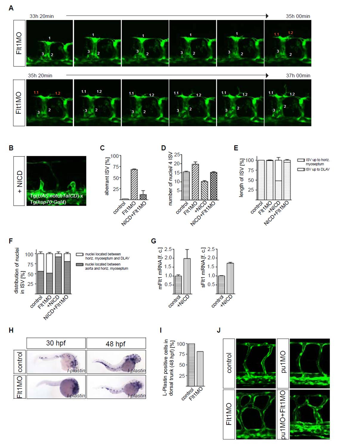
Fig. S3 Notch signaling and macrophages in the vascular phenotype of flt1 morpants. (A) Cell tracking in flt1 morphants shows that the endothelial cell leading the segmental sprout gives rise to two progeny cells that display tip cell filopodia characteristics. Embryos were incubated at 26.5°C during confocal imaging. (B) Overexpression of notch1a intracellular cleaved domain (NICD) in control embryos resulted in short segmental artery sprouts. (C) Statistical analysis of segmental vessel development in control (left), flt1 morphants (middle), and flt1 morphants after conditional overexpression of NICD (right) at 48 hpf. Note that overexpression of NICD restored segmental branching in flt1 morphants (right). (D,E) Quantification of endothelial nuclei per four ISV vessel sprouts and segmental sprout length. (F) Distribution of nuclei in the vessel sprout. Note that the distribution of nuclei is not restored in flt1 morphants after overexpression of NICD. (G) Taqman analysis of mflt1 and sflt1 after overexpression of NICD in embryos at 30 hpf. (H,I) Whole-mount in situ hybridization with the macrophage marker l-plastin and quantification of macrophage number. Note the overall reduced expression domain of l-plastin in flt1 morphants compared with controls. At 30 hpf, in flt1 morphants the trunk is almost devoid of l-plastin staining, suggesting that macrophages failed to migrate into this area. (J) Inhibiting macrophage formation by knockdown of pu1 did not affect segmental artery branching. Injection of pu1MO and Flt1MO together did not rescue the vascular branching phenotype. Error bars indicate s.e.m.
Image
Figure Caption
Acknowledgments
This image is the copyrighted work of the attributed author or publisher, and
ZFIN has permission only to display this image to its users.
Additional permissions should be obtained from the applicable author or publisher of the image.
Full text @ Development

