
Fig. 5
|
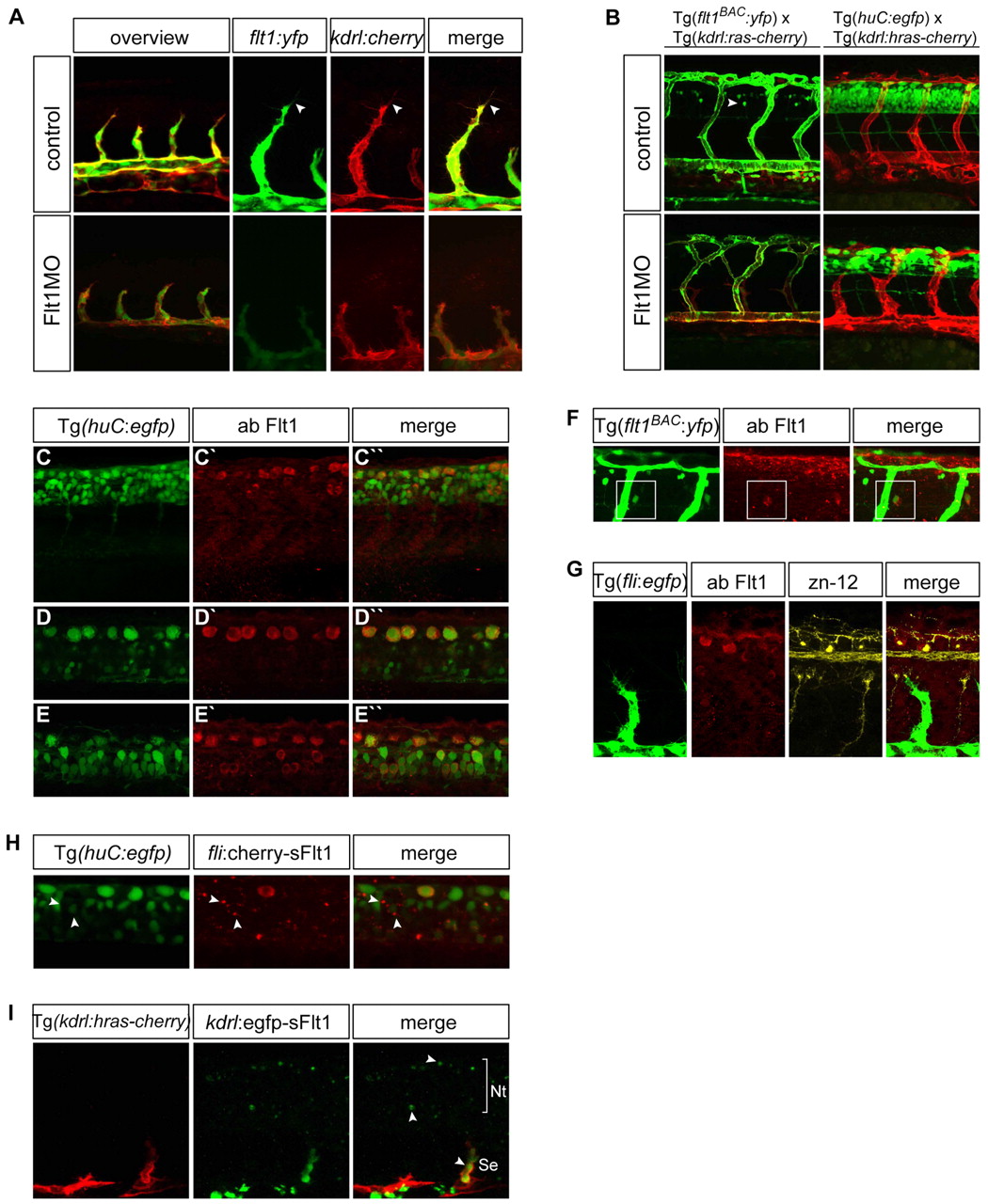
Fig. 5 Distribution of Flt1 in vessels and nerves. (A) Comparison of flt1BAC:yfp and kdrl:cherry expression in segmental artery sprouts of 30 hpf Tg(kdrl:ras-cherry)s916 � Tg(flt1BAC:yfp) double-transgenic zebrafish embryos, in controls (top row) and flt1 morphants (bottom row). In controls, note the overlap of flt1BAC:yfp and kdrl:cherry expression in tip and stalk cells. At 30 hpf, expression of flt1BAC:yfp is restricted to the vascular compartment. After injection of flt1 ATG-blocking MO, expression of flt1BAC:yfp in sprouts appears less intense, but is not completely lost. (B) Injection of flt1 ATG-blocking MO in Tg(kdrl:ras-cherry)s916 � Tg(flt1BAC:yfp) double-transgenic embryos causes hyperbranching of segmental vessels and affects neurons. In controls at 48 hpf, flt1BAC:yfp marks segmental vessels and a subset of spinal cord neurons (arrowhead). Injection of the flt1 ATG-blocking MO induces aberrant vessel patterning and the neuronal flt1BAC:yfp expression domain is reduced. Injection of the flt1 ATG-blocking MO in Tg(kdrl:hras-mcherry)s896 � Tg(huC:egfp) double-transgenic embryos affects neurons. (C-E3) Flt1 immunostaining in Tg(huC:egfp) neuronal reporter embryos shows Flt1 throughout the neural tube. Overview of neural tube (C-C3) and different focal planes from dorsal to ventral (D-E3) are shown. (F) Flt1 immunostaining in Tg(flt1BAC:yfp) embryos shows co-localization of flt1BAC:yfp with Flt1 antibody staining in spinal cord neurons (boxed). (G) Immunostaining for Flt1 and zn-12 shows Flt1 staining of Rohon-Beard sensory neurons. (H) Overexpression of fli1ep:cherry-sflt1 in Tg(huC:egfp) embryos results in neuronal sFlt1-cherry expression throughout the neural tube. Arrowheads indicate co-localization of huC:egfp with sFlt1-cherry on neuronal cell bodies. (I) Overexpression of kdrl:egfp-sflt1 in Tg(kdrl:hras-mcherry)s896 embryos shows sFlt1-egfp expression in vessels and the neural tube (arrowheads). Nt, neural tube; Se, segmental artery.