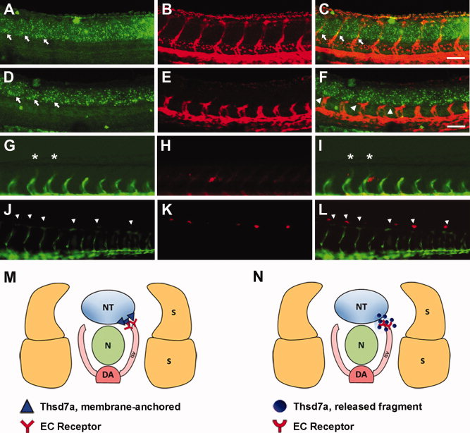
Fig. 5 thsd7a expression is associated with ISV development. A?F: Expression of thsd7a transcript (green) was detected by fluorescent ISH, followed by immunostaining to reveal the ISV pattern (red). Anterior is to the left. Expression of thsd7a in the ventral edge of neural tube is indicated by white arrows. Lateral deviation is indicated by white arrowheads. Scale bars represent 50 μm. Representative images showing the control embryo (A?C), and morphant at 30 hours postfertilization (hpf; D?F). G?L: Transplanted wild-type cells rescue the ISV phenotype. Embryos were examined at 28 hpf (G?I) and 50 hpf (J?L). ISVs were shown in green (G,J). Clusters of dextran-labeled cells were shown in red (H,K). G: ISVs that reached the dorsal region of the embryo were indicated by asterisk. H: Clusters of dextran-labeled cells were found in the recipients. I: Merged image of panel G and H. J,K: By 50 hpf, arrowheads indicate the normal ISVs that fused adjacently at the dorsal region of the embryo. Dextran-labeled cells were found associated with the normal ISVs. L: Images were merged. M,N: Models for the role of Thsd7a in EC migration during ISV angiogenesis. M: Thsd7a may be a membrane-anchored protein on the surface of neural cells, which distribute predominantly along the ventral edge of neural tube and located on the growth path of angiogenic ISVs. Interaction between Thsd7a and EC receptors directs EC migration during ISV angiogenesis. N: Fragments of Thsd7a may be released from neural cells, and form a concentration gradient that guide EC migration during ISV angiogenesis. NT, neural tube; N, notochord; DA, dorsal aorta; ISV, intersegmental vessel; S, somite.
Image
Figure Caption
Figure Data
Acknowledgments
This image is the copyrighted work of the attributed author or publisher, and
ZFIN has permission only to display this image to its users.
Additional permissions should be obtained from the applicable author or publisher of the image.
Full text @ Dev. Dyn.

